Copyright
©The Author(s) 2022.
World J Clin Cases. Sep 16, 2022; 10(26): 9510-9517
Published online Sep 16, 2022. doi: 10.12998/wjcc.v10.i26.9510
Published online Sep 16, 2022. doi: 10.12998/wjcc.v10.i26.9510
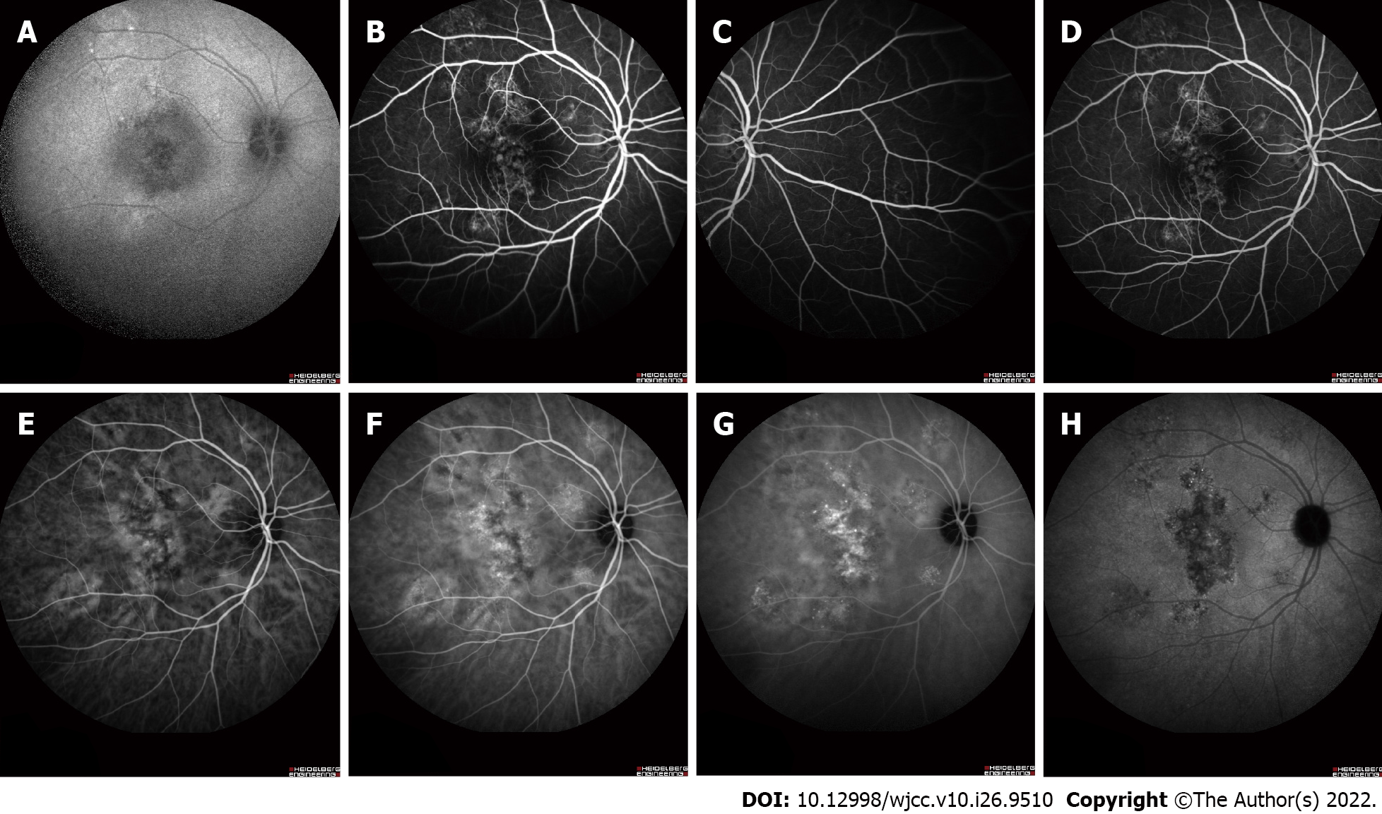
Figure 3 Fundus fluorescein angiography and indocyanine green angiography revealing some areas of transmission hyperfluorescence and dilated choroidal vessels of the right eye.
A: Fundus autofluorescence; B-D: Middle and late fundus fluorescein angiography; E-H: Early and late indocyanine green angiography.
- Citation: Xiang XL, Cao YH, Jiang TW, Huang ZR. Intravitreous injection of conbercept for bullous retinal detachment: A case report. World J Clin Cases 2022; 10(26): 9510-9517
- URL: https://www.wjgnet.com/2307-8960/full/v10/i26/9510.htm
- DOI: https://dx.doi.org/10.12998/wjcc.v10.i26.9510


