Copyright
©The Author(s) 2022.
World J Clin Cases. Sep 16, 2022; 10(26): 9417-9427
Published online Sep 16, 2022. doi: 10.12998/wjcc.v10.i26.9417
Published online Sep 16, 2022. doi: 10.12998/wjcc.v10.i26.9417
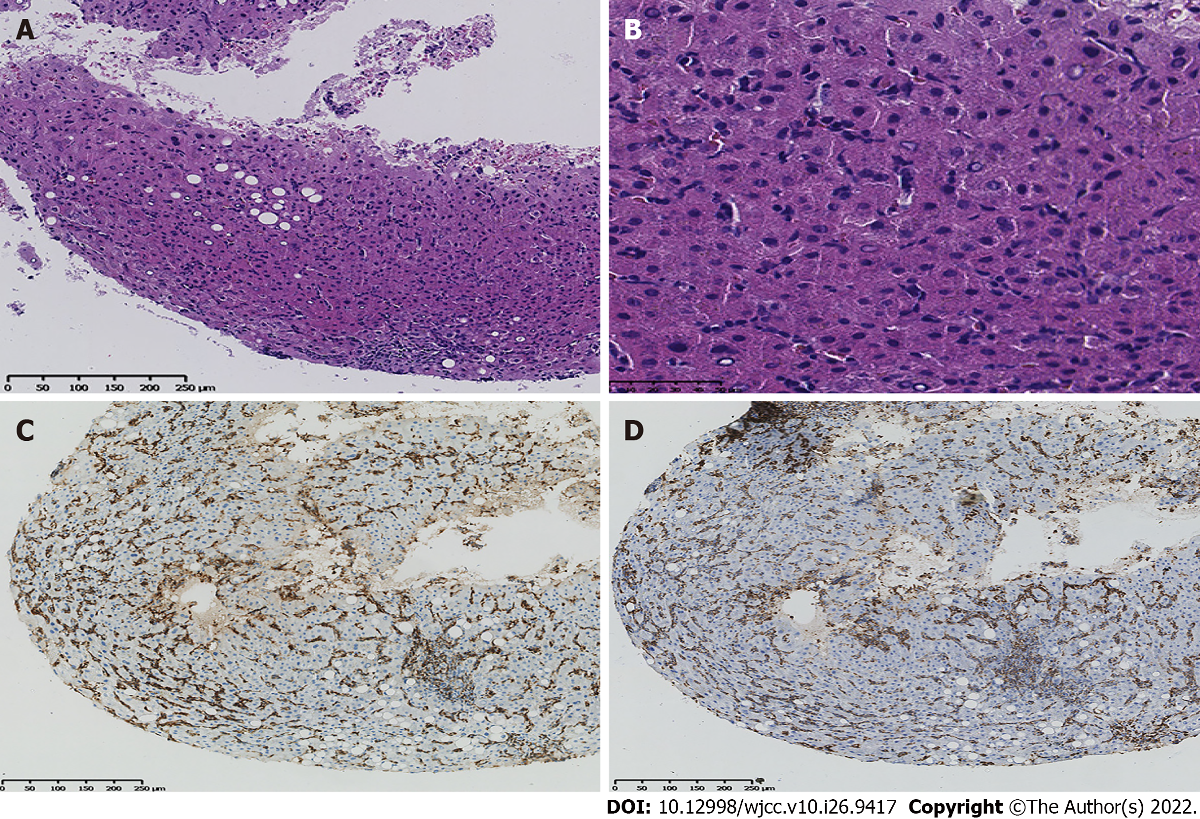
Figure 4 Pathology and immunochemistry were performed on four pieces of liver tissue.
A: Histopathological examination by haematoxylin and eosin staining (100 ×); B: Histopathological examination by haematoxylin and eosin staining (400×) showed few hepatocytes that were watery and lipomatous and multiple small focal lymphoid cell infiltrates; C: Immunochemical staining shows CD3 positivity; D: Immunochemical staining shows CD7 positivity. H&E, haematoxylin and eosin.
- Citation: Wu MM, Fu WJ, Wu J, Zhu LL, Niu T, Yang R, Yao J, Lu Q, Liao XY. Noncirrhotic portal hypertension due to peripheral T-cell lymphoma, not otherwise specified: A case report. World J Clin Cases 2022; 10(26): 9417-9427
- URL: https://www.wjgnet.com/2307-8960/full/v10/i26/9417.htm
- DOI: https://dx.doi.org/10.12998/wjcc.v10.i26.9417


