Copyright
©The Author(s) 2022.
World J Clin Cases. Sep 16, 2022; 10(26): 9417-9427
Published online Sep 16, 2022. doi: 10.12998/wjcc.v10.i26.9417
Published online Sep 16, 2022. doi: 10.12998/wjcc.v10.i26.9417
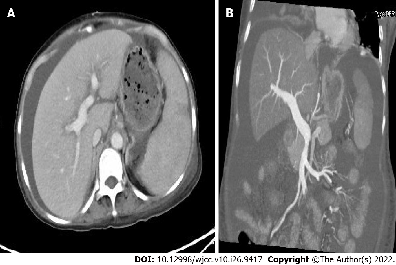
Figure 3 Liver computed tomographic arteriography.
A: Spleen growth: spleen enhancement was less uniform, and patchy slightly hypointense areas were seen, not excluding infarcts or intrahepatic lymphatic stasis; B: The main portal vein and splenic vein were slightly thickened, the diameter of the main portal vein was approximately 1.5 cm, and there was no thrombus or collateral circulation opening, A and B: The vessel was smooth and unobstructed.
- Citation: Wu MM, Fu WJ, Wu J, Zhu LL, Niu T, Yang R, Yao J, Lu Q, Liao XY. Noncirrhotic portal hypertension due to peripheral T-cell lymphoma, not otherwise specified: A case report. World J Clin Cases 2022; 10(26): 9417-9427
- URL: https://www.wjgnet.com/2307-8960/full/v10/i26/9417.htm
- DOI: https://dx.doi.org/10.12998/wjcc.v10.i26.9417


