Copyright
©The Author(s) 2022.
World J Clin Cases. Sep 16, 2022; 10(26): 9417-9427
Published online Sep 16, 2022. doi: 10.12998/wjcc.v10.i26.9417
Published online Sep 16, 2022. doi: 10.12998/wjcc.v10.i26.9417
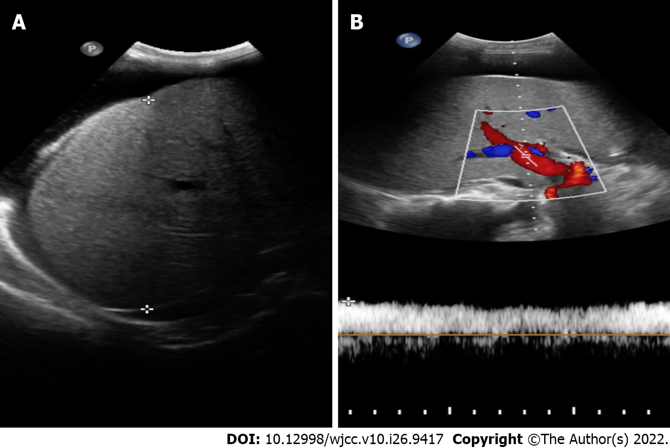
Figure 2 Liver ultrasound.
A: The liver capsule was less smooth, and the right liver had a maximum oblique transaxial distance of 15 cm and increased parenchymal echogenicity. The liver parenchyma was slightly thickened and heterogeneous without a definite space-occupying lesion; B: The diameter of the extrahepatic portal vein was approximately 12 mm, and the blood flow was unidirectional to the liver at a flow rate of 29.3 cm/s. The cava caliber and lumen appeared normal, as did blood flow in the hepatic vein, superior mesenteric vein, splenic vein, and inferior vena cava.
- Citation: Wu MM, Fu WJ, Wu J, Zhu LL, Niu T, Yang R, Yao J, Lu Q, Liao XY. Noncirrhotic portal hypertension due to peripheral T-cell lymphoma, not otherwise specified: A case report. World J Clin Cases 2022; 10(26): 9417-9427
- URL: https://www.wjgnet.com/2307-8960/full/v10/i26/9417.htm
- DOI: https://dx.doi.org/10.12998/wjcc.v10.i26.9417


