Copyright
©The Author(s) 2022.
World J Clin Cases. Sep 16, 2022; 10(26): 9340-9347
Published online Sep 16, 2022. doi: 10.12998/wjcc.v10.i26.9340
Published online Sep 16, 2022. doi: 10.12998/wjcc.v10.i26.9340
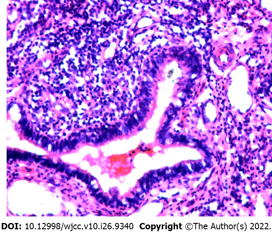
Figure 5 Pathology examination: Ciliated columnar epithelium, cartilage, and squamous cells lining the wall of the dilated, duct-like, cystic structure.
Obsolete hemorrhage and focal hyperplasia in the interstitial tissue are seen.
- Citation: Jin HJ, Yu Y, He W, Han Y. Posterior mediastinal extralobar pulmonary sequestration misdiagnosed as a neurogenic tumor: A case report. World J Clin Cases 2022; 10(26): 9340-9347
- URL: https://www.wjgnet.com/2307-8960/full/v10/i26/9340.htm
- DOI: https://dx.doi.org/10.12998/wjcc.v10.i26.9340


