Copyright
©The Author(s) 2022.
World J Clin Cases. Sep 6, 2022; 10(25): 9148-9155
Published online Sep 6, 2022. doi: 10.12998/wjcc.v10.i25.9148
Published online Sep 6, 2022. doi: 10.12998/wjcc.v10.i25.9148
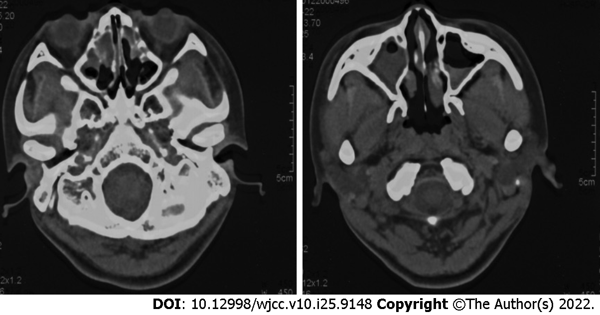
Figure 2 Computed tomography image of the sinus showing the mucosa of bilateral ethmoid sinus and maxillary sinus was thickened and edematous, and the lesion of the right sinus cavity was more serious than that of the left.
- Citation: Zhang YY, Lou Y, Yan H, Tang H. CCNO mutation as a cause of primary ciliary dyskinesia: A case report. World J Clin Cases 2022; 10(25): 9148-9155
- URL: https://www.wjgnet.com/2307-8960/full/v10/i25/9148.htm
- DOI: https://dx.doi.org/10.12998/wjcc.v10.i25.9148


