Copyright
©The Author(s) 2022.
World J Clin Cases. Sep 6, 2022; 10(25): 9104-9111
Published online Sep 6, 2022. doi: 10.12998/wjcc.v10.i25.9104
Published online Sep 6, 2022. doi: 10.12998/wjcc.v10.i25.9104
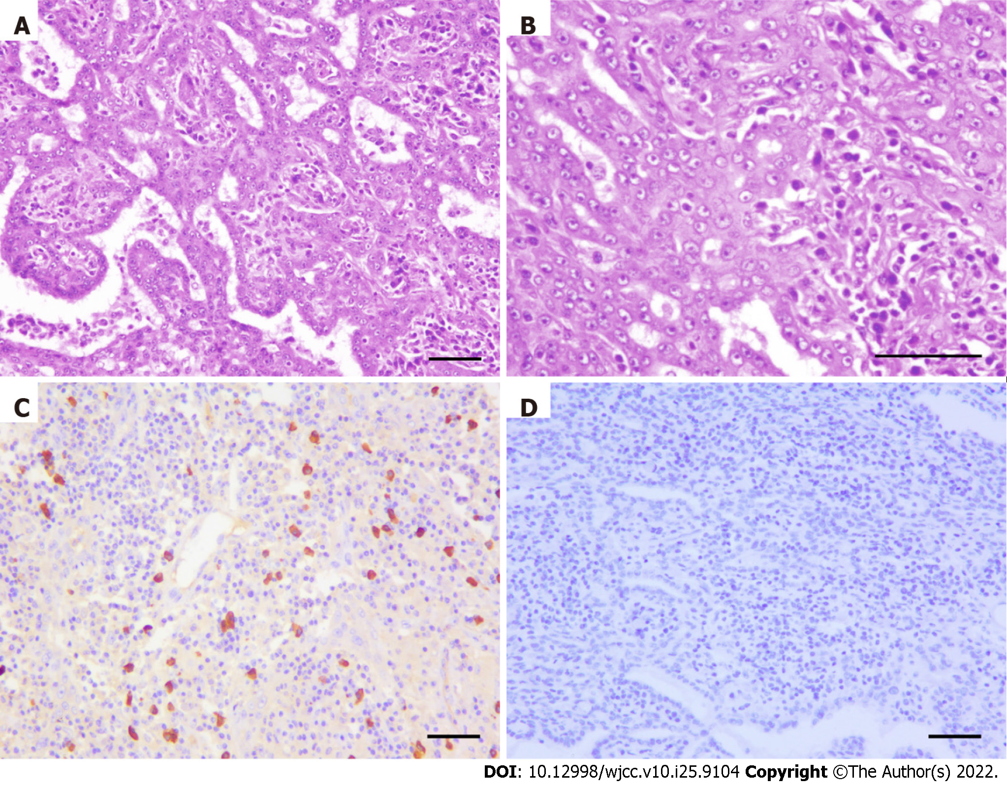
Figure 4 Microscopic pathology of surgical specimens.
A and B: H & E staining of hepatic lesion, (A) H & E × 100 (B) H & E × 200. The malignant component of a tumor consists of deformed fused glandular ducts that form a sieve, and cord-like structures. The neoplastic cells are of medium size with well-defined nucleoli, and most of the cytoplasm is pale, slightly acidophilic or vacuolated; C and D: Immunohistochemical staining of IgG4 and HHV8 in mediastinal lymph node (× 100). Bar = 100 μm.
- Citation: Wang SC, Chen YY, Cheng F, Wang HY, Wu FS, Teng LS. Malignant transformation of biliary adenofibroma combined with benign lymphadenopathy mimicking advanced liver carcinoma: A case report. World J Clin Cases 2022; 10(25): 9104-9111
- URL: https://www.wjgnet.com/2307-8960/full/v10/i25/9104.htm
- DOI: https://dx.doi.org/10.12998/wjcc.v10.i25.9104


