Copyright
©The Author(s) 2022.
World J Clin Cases. Sep 6, 2022; 10(25): 9104-9111
Published online Sep 6, 2022. doi: 10.12998/wjcc.v10.i25.9104
Published online Sep 6, 2022. doi: 10.12998/wjcc.v10.i25.9104
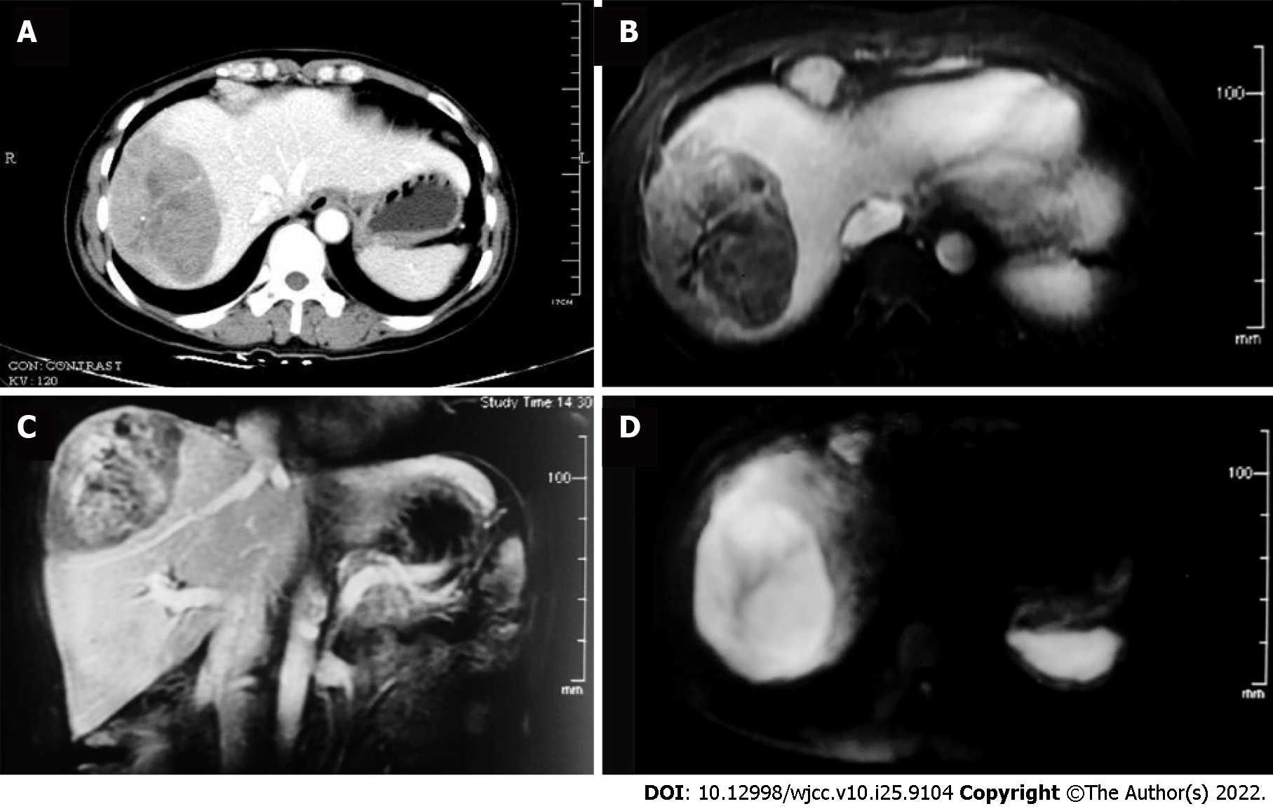
Figure 1 Computed tomography, magnetic resonance imaging and positron emission tomography/computed tomography images of liver mass and mediastinal lymphadenopathy.
A: Abdominal contrast computed tomography, venous phase; B-D: Representative images from the MRI study (B: Venous phase; C: Sagittal venous phase; D: Diffusion weighted).
- Citation: Wang SC, Chen YY, Cheng F, Wang HY, Wu FS, Teng LS. Malignant transformation of biliary adenofibroma combined with benign lymphadenopathy mimicking advanced liver carcinoma: A case report. World J Clin Cases 2022; 10(25): 9104-9111
- URL: https://www.wjgnet.com/2307-8960/full/v10/i25/9104.htm
- DOI: https://dx.doi.org/10.12998/wjcc.v10.i25.9104


