Copyright
©The Author(s) 2022.
World J Clin Cases. Sep 6, 2022; 10(25): 9096-9103
Published online Sep 6, 2022. doi: 10.12998/wjcc.v10.i25.9096
Published online Sep 6, 2022. doi: 10.12998/wjcc.v10.i25.9096
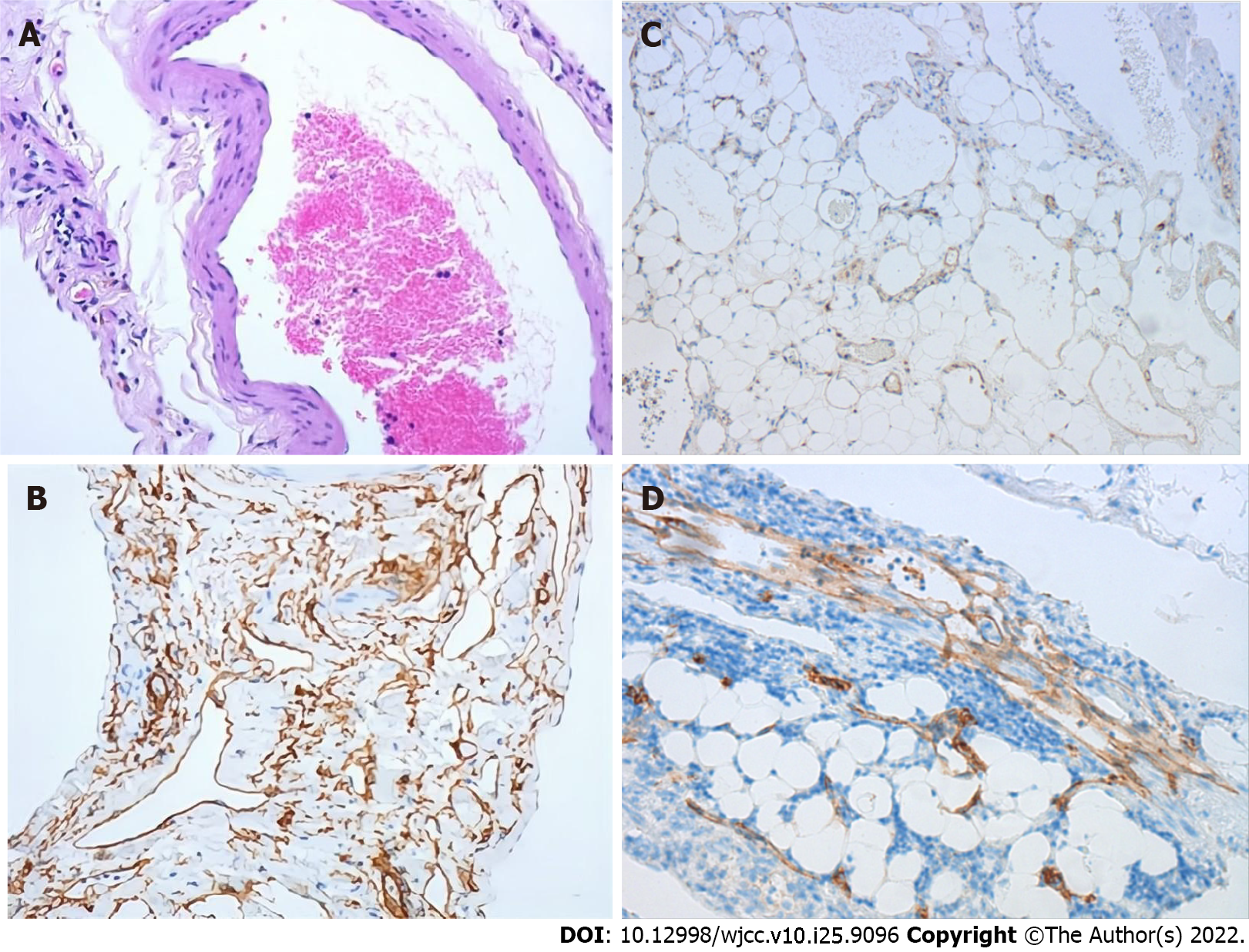
Figure 3 Pathologic findings of the venous hemangioma.
A: The specimen tissues are composed of various-sized cystic cavities with walls consisting of single-layered flattened cells; B: Immunohistochemically, the endothelial cells are positive for CD34; C: Some endothelial cells are positive for CD31; D: Some endothelial cells are positive for D2-40.
- Citation: Qin Y, Qiao P, Guan X, Zeng S, Hu XP, Wang B. Successful resection of a huge retroperitoneal venous hemangioma: A case report. World J Clin Cases 2022; 10(25): 9096-9103
- URL: https://www.wjgnet.com/2307-8960/full/v10/i25/9096.htm
- DOI: https://dx.doi.org/10.12998/wjcc.v10.i25.9096


