Copyright
©The Author(s) 2022.
World J Clin Cases. Sep 6, 2022; 10(25): 9096-9103
Published online Sep 6, 2022. doi: 10.12998/wjcc.v10.i25.9096
Published online Sep 6, 2022. doi: 10.12998/wjcc.v10.i25.9096
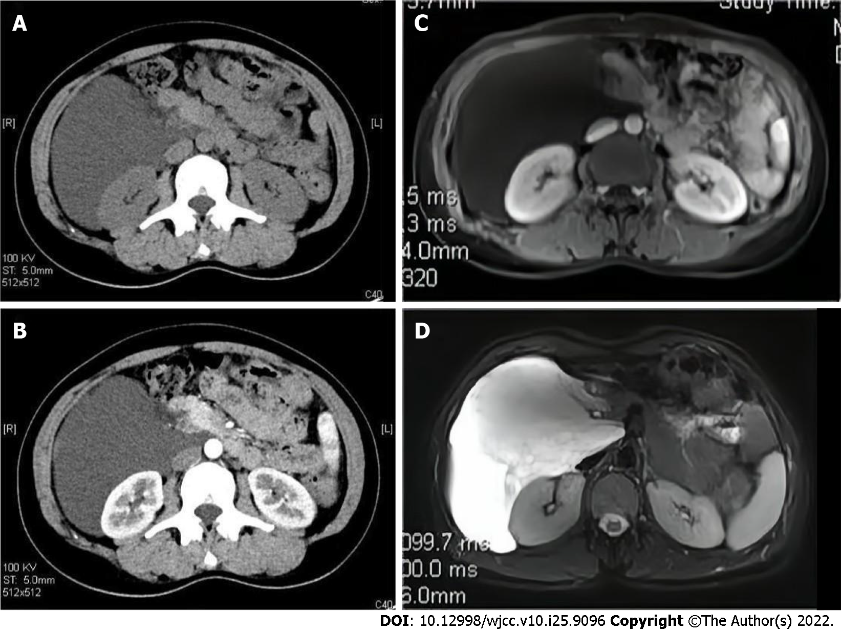
Figure 1 Imaging findings of a retroperitoneal mass.
A: CT scans of the abdomen demonstrates a hypodense mass in the right retroperitoneum; B: The enhanced CT shows the mass without remarkable enhancement; C: On MRI, T1-weighted image shows the mass as low signal intensity without enhancement; D: T2-weighted image shows the mass with high signal intensity. CT: Computed tomography; MRI: Magnetic resonance imaging.
- Citation: Qin Y, Qiao P, Guan X, Zeng S, Hu XP, Wang B. Successful resection of a huge retroperitoneal venous hemangioma: A case report. World J Clin Cases 2022; 10(25): 9096-9103
- URL: https://www.wjgnet.com/2307-8960/full/v10/i25/9096.htm
- DOI: https://dx.doi.org/10.12998/wjcc.v10.i25.9096


