Copyright
©The Author(s) 2022.
World J Clin Cases. Sep 6, 2022; 10(25): 9050-9056
Published online Sep 6, 2022. doi: 10.12998/wjcc.v10.i25.9050
Published online Sep 6, 2022. doi: 10.12998/wjcc.v10.i25.9050
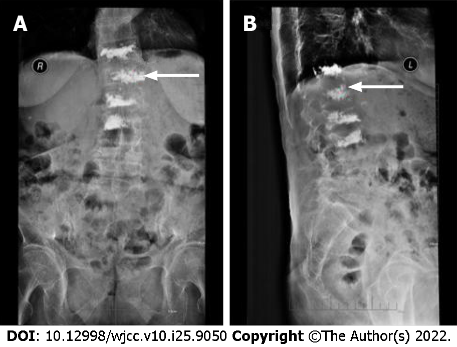
Figure 2 Postoperative X-ray.
A: Postoperative anteroposterior X-ray showed an almost evenly distributed cement of L1; B: Postoperative lateral X-ray showed a safe placement of the cement (white arrows).
- Citation: Wang YF, Bian ZY, Li XX, Hu YX, Jiang L. Total spinal anesthesia caused by lidocaine during unilateral percutaneous vertebroplasty performed under local anesthesia: A case report. World J Clin Cases 2022; 10(25): 9050-9056
- URL: https://www.wjgnet.com/2307-8960/full/v10/i25/9050.htm
- DOI: https://dx.doi.org/10.12998/wjcc.v10.i25.9050


