Copyright
©The Author(s) 2022.
World J Clin Cases. Sep 6, 2022; 10(25): 9044-9049
Published online Sep 6, 2022. doi: 10.12998/wjcc.v10.i25.9044
Published online Sep 6, 2022. doi: 10.12998/wjcc.v10.i25.9044
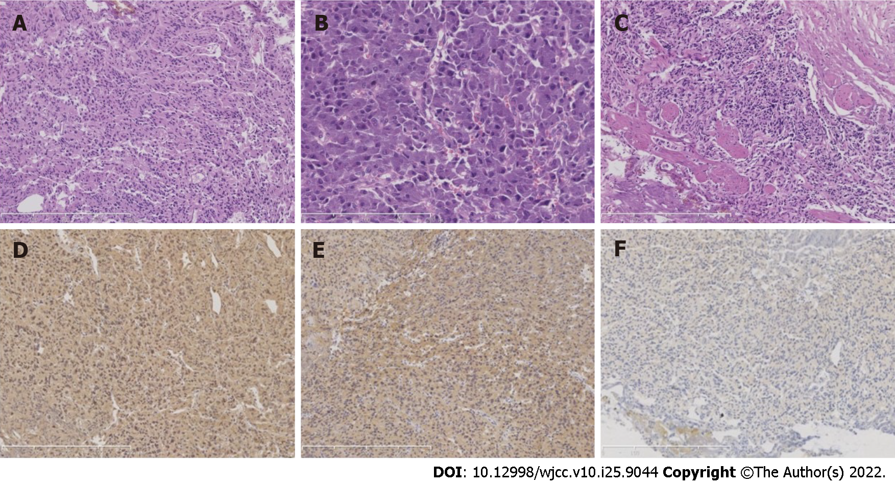
Figure 2 Histological examination and immunohistochemical staining.
A: Histological examination of the resected vesical polyp revealed that the cells were arranged in sheets and nests [hematoxylin and eosin (H&E) staining]; B: The cytoplasm was eosinophilic to amphophilic (H&E staining); C: The neoplasm infiltrated the muscle layer (H&E staining); D-F: Homogeneous immunoreactivity to neuroendocrine markers such as synaptophysin (D), chromogranin A (E), and S-100 (F) (immunohistochemical staining).
- Citation: Wang L, Zhang YN, Chen GY. Bladder paraganglioma after kidney transplantation: A case report. World J Clin Cases 2022; 10(25): 9044-9049
- URL: https://www.wjgnet.com/2307-8960/full/v10/i25/9044.htm
- DOI: https://dx.doi.org/10.12998/wjcc.v10.i25.9044


