Copyright
©The Author(s) 2022.
World J Clin Cases. Sep 6, 2022; 10(25): 9020-9027
Published online Sep 6, 2022. doi: 10.12998/wjcc.v10.i25.9020
Published online Sep 6, 2022. doi: 10.12998/wjcc.v10.i25.9020
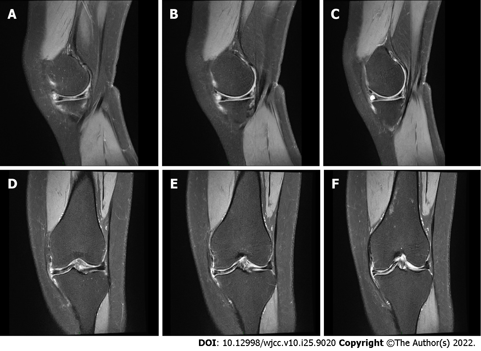
Figure 3 Magnetic resonance imaging examination of the left knee.
A-C: Three consecutive images in the sagittal view show a complete medial discoid meniscus; D-F: Coronal views showing a horizontal tear of the medial meniscus.
- Citation: Zheng ZR, Ma H, Yang F, Yuan L, Wang GD, Zhao XW, Ma LF. Discoid medial meniscus of both knees: A case report . World J Clin Cases 2022; 10(25): 9020-9027
- URL: https://www.wjgnet.com/2307-8960/full/v10/i25/9020.htm
- DOI: https://dx.doi.org/10.12998/wjcc.v10.i25.9020


