Copyright
©The Author(s) 2022.
World J Clin Cases. Sep 6, 2022; 10(25): 9012-9019
Published online Sep 6, 2022. doi: 10.12998/wjcc.v10.i25.9012
Published online Sep 6, 2022. doi: 10.12998/wjcc.v10.i25.9012
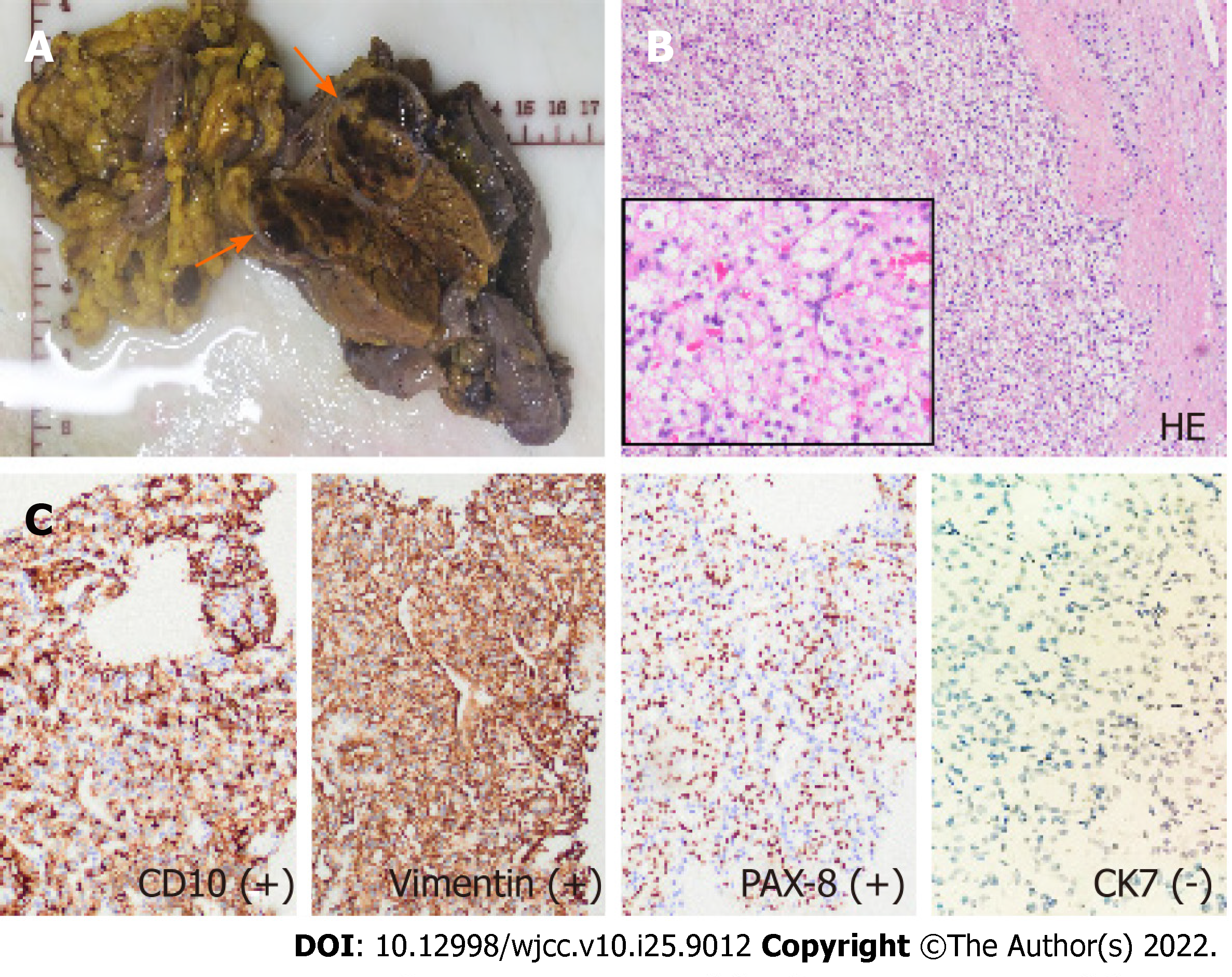
Figure 4 Postoperative histopathology of the resected specimen.
A: The pancreatic mass shows hemorrhagic and necrotic changes; there is a thin fibrous capsule (arrow); B: Stained section shows large polygonal cells with clear cytoplasm, arranged in an alveolar pattern; the cells have uniform round nuclei with inconspicuous nucleoli (hematoxylin and eosin, × 100, inset × 400); and C: Immunohistochemistry shows the tumor cells to be positive for vimentin, CD10, and PAX-8, and negative for CK7.
- Citation: Liang XK, Li LJ, He YM, Xu ZF. Misdiagnosis of pancreatic metastasis from renal cell carcinoma: A case report. World J Clin Cases 2022; 10(25): 9012-9019
- URL: https://www.wjgnet.com/2307-8960/full/v10/i25/9012.htm
- DOI: https://dx.doi.org/10.12998/wjcc.v10.i25.9012


