Copyright
©The Author(s) 2022.
World J Clin Cases. Sep 6, 2022; 10(25): 9012-9019
Published online Sep 6, 2022. doi: 10.12998/wjcc.v10.i25.9012
Published online Sep 6, 2022. doi: 10.12998/wjcc.v10.i25.9012
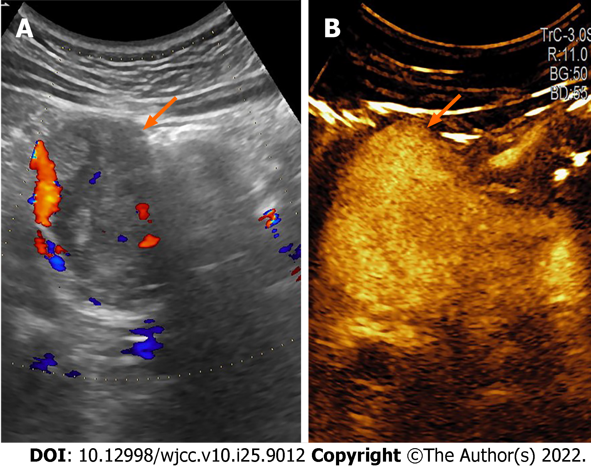
Figure 1 Sonogram imagine.
A: B-mode; B: Contrast-enhanced ultrasound. There is a hypoechoic lesion (arrow) in the pancreatic head, with high and inhomogeneous enhancement.
- Citation: Liang XK, Li LJ, He YM, Xu ZF. Misdiagnosis of pancreatic metastasis from renal cell carcinoma: A case report. World J Clin Cases 2022; 10(25): 9012-9019
- URL: https://www.wjgnet.com/2307-8960/full/v10/i25/9012.htm
- DOI: https://dx.doi.org/10.12998/wjcc.v10.i25.9012


