Copyright
©The Author(s) 2022.
World J Clin Cases. Sep 6, 2022; 10(25): 8968-8973
Published online Sep 6, 2022. doi: 10.12998/wjcc.v10.i25.8968
Published online Sep 6, 2022. doi: 10.12998/wjcc.v10.i25.8968
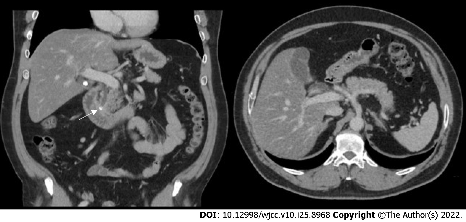
Figure 2 Contrast-enhanced computed tomography image.
Contrast-enhanced computed tomography scan present hyperdense gallstone and previous cystic duct stone become lodged in the common bile duct (arrow). Recovery of splenic infarction.
- Citation: Wu CY, Su CC, Huang HH, Wang YT, Wang CC. Gallstone associated celiac trunk thromboembolisms complicated with splenic infarction: A case report. World J Clin Cases 2022; 10(25): 8968-8973
- URL: https://www.wjgnet.com/2307-8960/full/v10/i25/8968.htm
- DOI: https://dx.doi.org/10.12998/wjcc.v10.i25.8968


