Copyright
©The Author(s) 2022.
World J Clin Cases. Sep 6, 2022; 10(25): 8968-8973
Published online Sep 6, 2022. doi: 10.12998/wjcc.v10.i25.8968
Published online Sep 6, 2022. doi: 10.12998/wjcc.v10.i25.8968
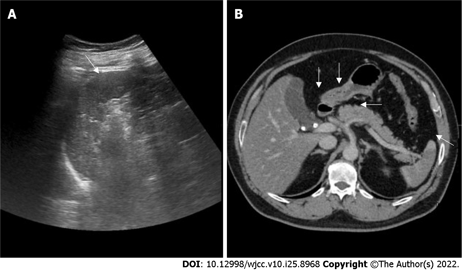
Figure 1 Ultrasound and contras-enhanced computed tomography images.
A: Ultrasound showed hypoechoic area compared to the rest of the spleen; B: Contras-enhanced computed tomography revealed hyperdense stones at gallbladder and cystic duct. Irregular filling defect in celiac trunk, common hepatic artery and partial splenic artery along with fat stranding, and focal wedge-shaped hypoenhancing region of spleen (arrows), suggesting extensive thrombosis following splenic infarction.
- Citation: Wu CY, Su CC, Huang HH, Wang YT, Wang CC. Gallstone associated celiac trunk thromboembolisms complicated with splenic infarction: A case report. World J Clin Cases 2022; 10(25): 8968-8973
- URL: https://www.wjgnet.com/2307-8960/full/v10/i25/8968.htm
- DOI: https://dx.doi.org/10.12998/wjcc.v10.i25.8968


