Copyright
©The Author(s) 2022.
World J Clin Cases. Sep 6, 2022; 10(25): 8954-8961
Published online Sep 6, 2022. doi: 10.12998/wjcc.v10.i25.8954
Published online Sep 6, 2022. doi: 10.12998/wjcc.v10.i25.8954
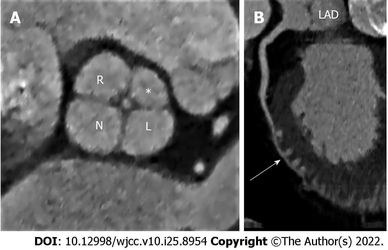
Figure 2 Multislice computed tomography image of the quadricuspid aortic valve.
A: Quadricuspid aortic valve depicted by multiplanar reformatted computed tomography (CT) image during diastole. The supranumerary cusp (asterisk) is located between the right and left coronary cusp. Central malcoaptation of the cusps can also be seen; B: Curved planar reformation of coronary CT angiography shows the right-ventricular type of myocardial bridging at the distal segment of the left anterior descending artery (marked by arrow). R: Right coronary cusp; L: Left coronary cusp; N: Noncoronary cusp; *: Supernumerary cusp; LAD: Left anterior descending.
- Citation: Sopek Merkaš I, Lakušić N, Paar MH. Quadricuspid aortic valve and right ventricular type of myocardial bridging in an asymptomatic middle-aged woman: A case report. World J Clin Cases 2022; 10(25): 8954-8961
- URL: https://www.wjgnet.com/2307-8960/full/v10/i25/8954.htm
- DOI: https://dx.doi.org/10.12998/wjcc.v10.i25.8954


