Copyright
©The Author(s) 2022.
World J Clin Cases. Sep 6, 2022; 10(25): 8939-8944
Published online Sep 6, 2022. doi: 10.12998/wjcc.v10.i25.8939
Published online Sep 6, 2022. doi: 10.12998/wjcc.v10.i25.8939
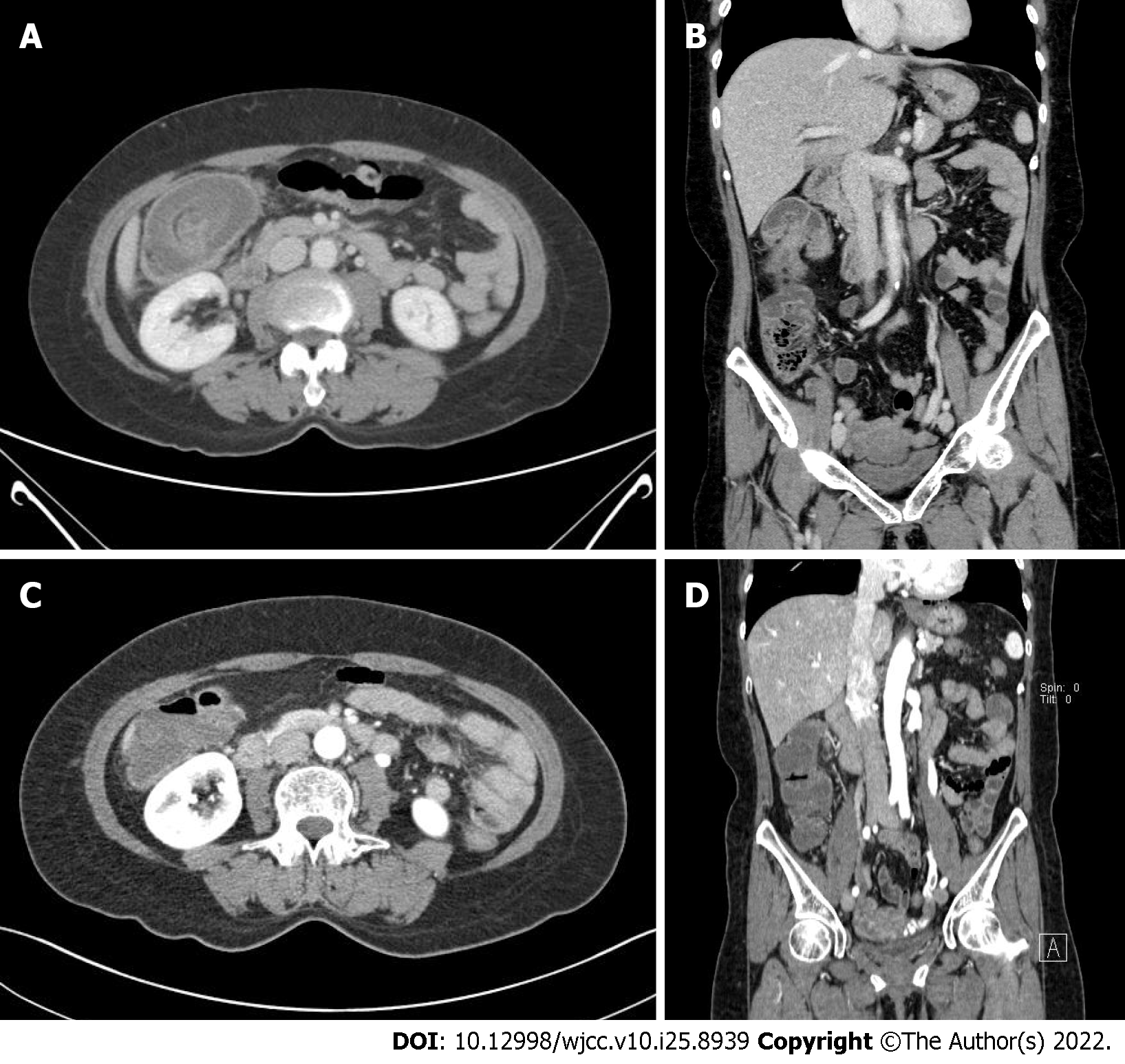
Figure 2 Initial and follow-up contrast-enhanced computed tomography.
A: Axial portal phase image shows a target-like lesion in the right side colon with bowel and fatty mesentery inside and colon wall thickening with submucosal swelling and highly attenuated infiltration of adjacent pericolic fat; B: Coronal portal phase image shows invagination of the right side colon; C: Axial portal phase shows resolved state of previously seen colon wall swelling and target-like lesion; D: Coronal portal phase image shows resolved state of previously seen invagination.
- Citation: Moon JY, Lee MR, Yim SK, Ha GW. Colo-colonic intussusception with post-polypectomy electrocoagulation syndrome: A case report. World J Clin Cases 2022; 10(25): 8939-8944
- URL: https://www.wjgnet.com/2307-8960/full/v10/i25/8939.htm
- DOI: https://dx.doi.org/10.12998/wjcc.v10.i25.8939


