Copyright
©The Author(s) 2022.
World J Clin Cases. Sep 6, 2022; 10(25): 8844-8853
Published online Sep 6, 2022. doi: 10.12998/wjcc.v10.i25.8844
Published online Sep 6, 2022. doi: 10.12998/wjcc.v10.i25.8844
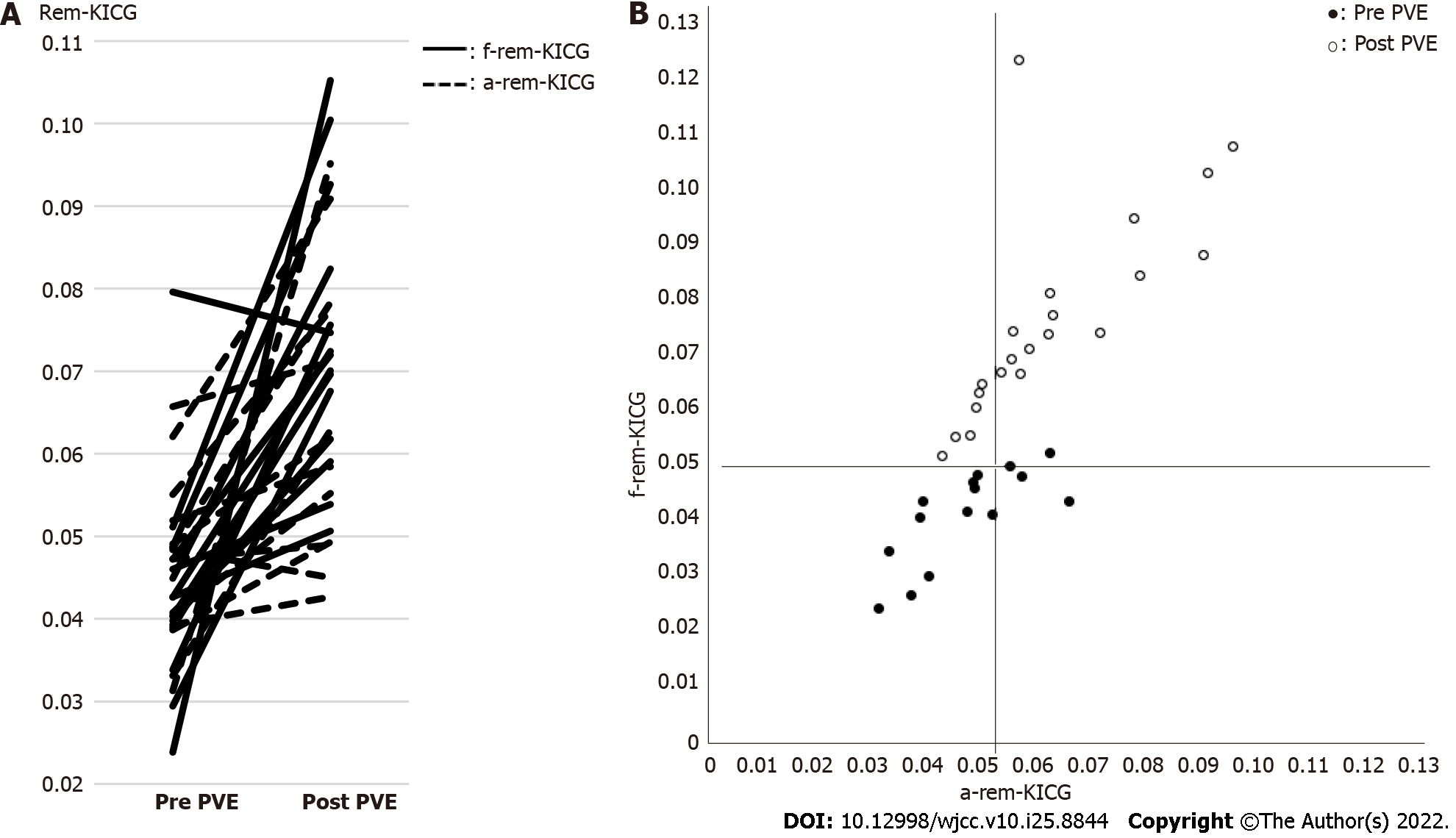
Figure 2 Anatomical volume remnant indocyanine green plasma clearance rate and functional volume remnant indocyanine green plasma clearance rate.
A: Changes in anatomical volume remnant indocyanine green plasma clearance rate (a-rem-KICG) and functional volume remnant indocyanine green plasma clearance rate (f-rem-KICG) between pre- and post-portal vein embolization. The increased amount of f-rem-KICG was significantly larger than that of a-rem-KICG (P = 0.0273); B: Scatter plots of a-rem-KICG and f-rem-KICG. The black and white dots show the pre-and post-portal vein embolization (PVE) remnant liver KICGs, respectively. After PVE, 17 patients had a-rem-KICG > 0.05, and f-rem-KICG > 0.05. Six patients (26%) had a-rem-KICG < 0.05, and f-rem-KICG > 0.05. A significant correlation was observed between a-rem-KICG and f-rem-KICG (P = 0.0002, R2 = 0.5156). a-rem-KICG: Anatomical volume remnant indocyanine green plasma clearance rate; f-rem-KICG: Functional volume remnant indocyanine green plasma clearance rate; PVE: Portal vein embolization.
- Citation: Iwaki K, Kaihara S, Kita R, Kitamura K, Hashida H, Uryuhara K. Indocyanine green plasma clearance rate and 99mTc-galactosyl human serum albumin single-photon emission computed tomography evaluated preoperative remnant liver. World J Clin Cases 2022; 10(25): 8844-8853
- URL: https://www.wjgnet.com/2307-8960/full/v10/i25/8844.htm
- DOI: https://dx.doi.org/10.12998/wjcc.v10.i25.8844


