Copyright
©The Author(s) 2022.
World J Clin Cases. Aug 26, 2022; 10(24): 8782-8787
Published online Aug 26, 2022. doi: 10.12998/wjcc.v10.i24.8782
Published online Aug 26, 2022. doi: 10.12998/wjcc.v10.i24.8782
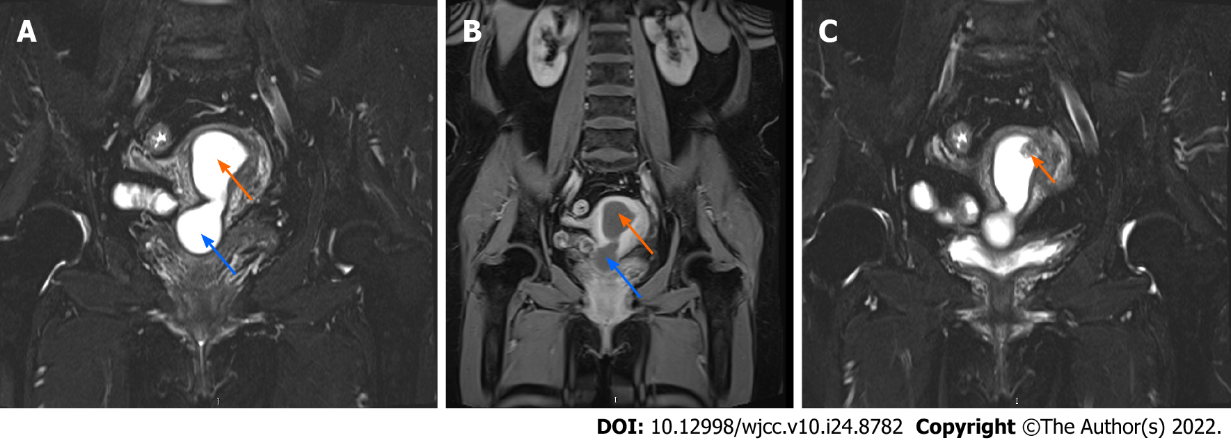
Figure 1 Magnetic resonance imaging of the uterine cavity.
A and B: T2 sequencing (A) and enhanced sequencing (B) of the uterus, both showing fluid within the uterine cavity (orange arrow) and the cervical canal (blue arrow), which was most likely pyometra; C: A mass that protruded into the uterine cavity (arrow), which is most likely an endometrial polyp or submucosal myoma.
- Citation: Shu XY, Dai Z, Zhang S, Yang HX, Bi H. Endometrial squamous cell carcinoma originating from the cervix: A case report. World J Clin Cases 2022; 10(24): 8782-8787
- URL: https://www.wjgnet.com/2307-8960/full/v10/i24/8782.htm
- DOI: https://dx.doi.org/10.12998/wjcc.v10.i24.8782


