Copyright
©The Author(s) 2022.
World J Clin Cases. Aug 26, 2022; 10(24): 8775-8781
Published online Aug 26, 2022. doi: 10.12998/wjcc.v10.i24.8775
Published online Aug 26, 2022. doi: 10.12998/wjcc.v10.i24.8775
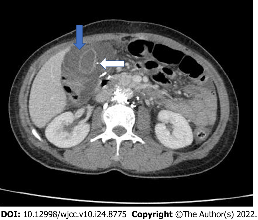
Figure 4 Contrast-enhanced abdominal computed tomography after 3 days.
Axial image shows further increased gallbladder wall thickening (~20 mm) and subserosal edema (blue arrow), without evidence of stones, pseudoaneurysm, or contrast agent leakage. A small, high-density nodule can be observed in the gallbladder wall (white arrow).
- Citation: Dung LV, Hien MM, Tra My TT, Luu DT, Linh LT, Duc NM. Cholecystitis-an uncommon complication following thoracic duct embolization for chylothorax: A case report. World J Clin Cases 2022; 10(24): 8775-8781
- URL: https://www.wjgnet.com/2307-8960/full/v10/i24/8775.htm
- DOI: https://dx.doi.org/10.12998/wjcc.v10.i24.8775


