Copyright
©The Author(s) 2022.
World J Clin Cases. Aug 26, 2022; 10(24): 8735-8741
Published online Aug 26, 2022. doi: 10.12998/wjcc.v10.i24.8735
Published online Aug 26, 2022. doi: 10.12998/wjcc.v10.i24.8735
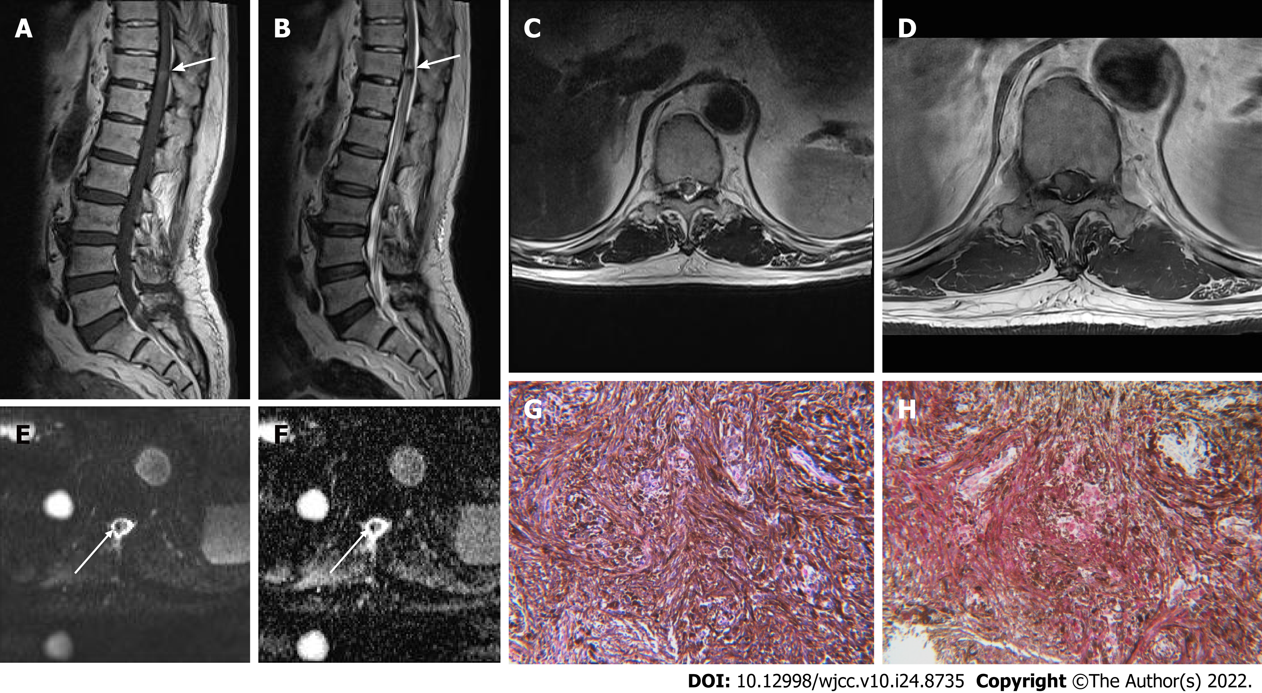
Figure 2 A 72-year-old male with non-psammomatous melanotic schwannoma located in the spinal cord.
A: Sagittal T1-weighted image of thoracolumbar spine shows a well-defined round-shaped nodular mass lesion (arrow) with increased signal intensity in the spinal cord of T11 level; B: The mass reveals dark signal intensity (arrow) such as a signal void on a sagittal T2-weighted image; C: On the corresponding axial T2-weighted image, high signal edema adjacent to dark signal intensity lesion (arrows) is noted in the spinal cord; D: The mass shows uniform homogenous enhancement (arrows) after gadolinium-contrast injection. It is eccentrically located on the right side within the distal spinal cord; E and F: On diffusion-weighted imaging and an apparent diffusion coefficient map, signal void (arrows) is noted within the mass without diffusion restriction; G: Section reveals a spindle cell lesion with dense melanin pigmentation that covers the nucleus and cytoplasm (hematoxylin and eosin, × 200); H: Immunostaining shows diffuse red staining for S100 protein (× 200).
- Citation: Yeom JA, Song YS, Lee IS, Han IH, Choi KU. Malignant melanotic nerve sheath tumors in the spinal canal of psammomatous and non-psammomatous type: Two case reports. World J Clin Cases 2022; 10(24): 8735-8741
- URL: https://www.wjgnet.com/2307-8960/full/v10/i24/8735.htm
- DOI: https://dx.doi.org/10.12998/wjcc.v10.i24.8735


