Copyright
©The Author(s) 2022.
World J Clin Cases. Aug 26, 2022; 10(24): 8735-8741
Published online Aug 26, 2022. doi: 10.12998/wjcc.v10.i24.8735
Published online Aug 26, 2022. doi: 10.12998/wjcc.v10.i24.8735
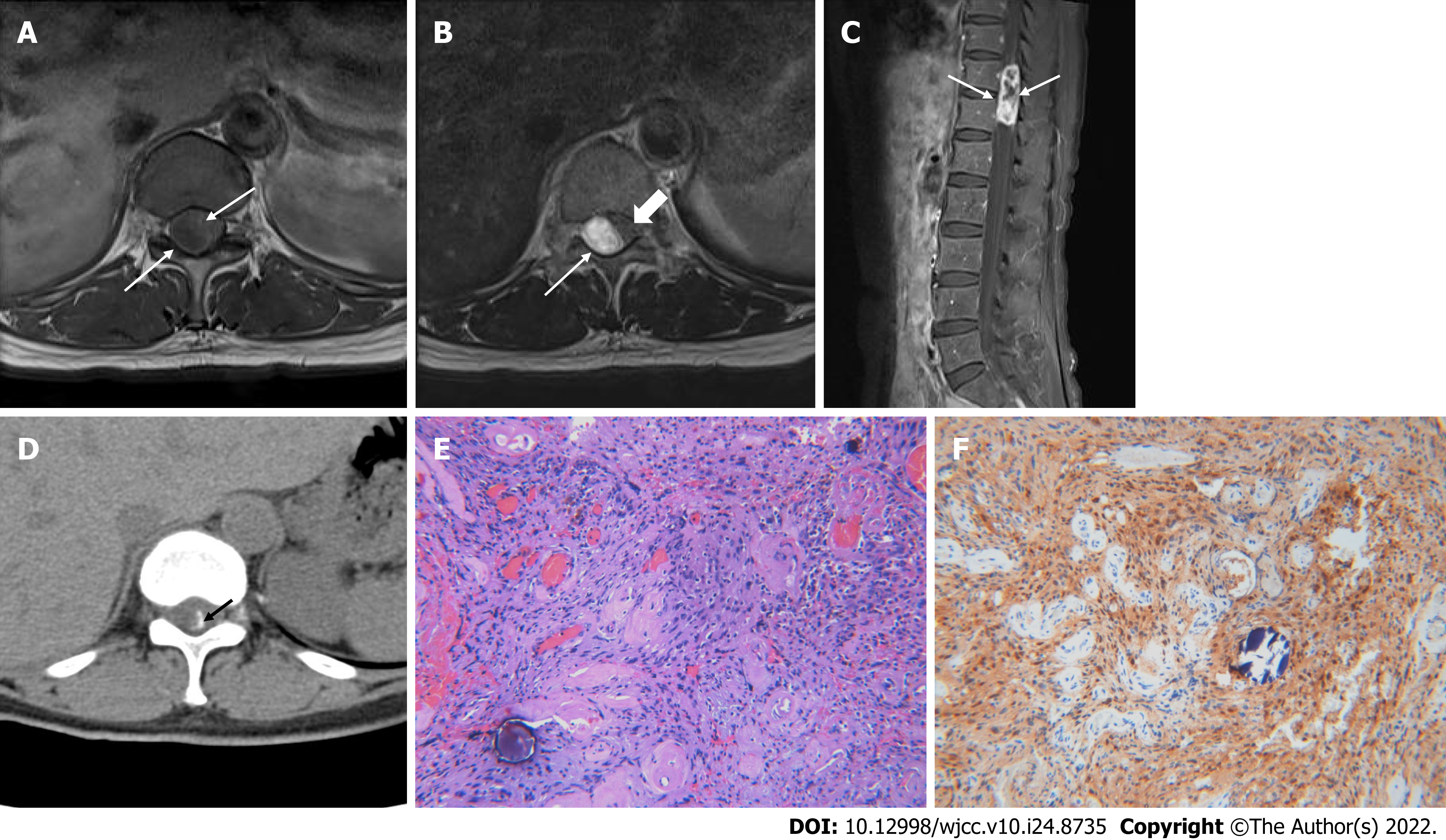
Figure 1 A 58-year-old female with psammomatous melanotic schwannoma.
A: Axial T1-weighted image of 11-12th thoracic spine level shows low signal mass lesion (arrows) located in the intradural space; B: Axial T2-weighted image shows the mass lesion (arrows) with heterogeneously high signal intensity and the spinal cord (thick arrow) is displaced and compressed by the mass lesion; C: The mass lesion (arrows) represents heterogeneously strong enhancement containing necrotic portion on sagittal fat-suppressed, contrast-enhanced T1-weighted image; D: Amorphous linear calcification (black arrow) is noted in the peripheral margin of the mass on the computed tomography scan; E: Section shows spindle-shaped Schwann cells with brownish pigments, psammoma bodies (hematoxylin and eosin, × 100); F: Positive immunoactivity for S-100 protein that are characteristic features of psammomatous melanotic schwannoma (× 100).
- Citation: Yeom JA, Song YS, Lee IS, Han IH, Choi KU. Malignant melanotic nerve sheath tumors in the spinal canal of psammomatous and non-psammomatous type: Two case reports. World J Clin Cases 2022; 10(24): 8735-8741
- URL: https://www.wjgnet.com/2307-8960/full/v10/i24/8735.htm
- DOI: https://dx.doi.org/10.12998/wjcc.v10.i24.8735


