Copyright
©The Author(s) 2022.
World J Clin Cases. Aug 26, 2022; 10(24): 8709-8717
Published online Aug 26, 2022. doi: 10.12998/wjcc.v10.i24.8709
Published online Aug 26, 2022. doi: 10.12998/wjcc.v10.i24.8709
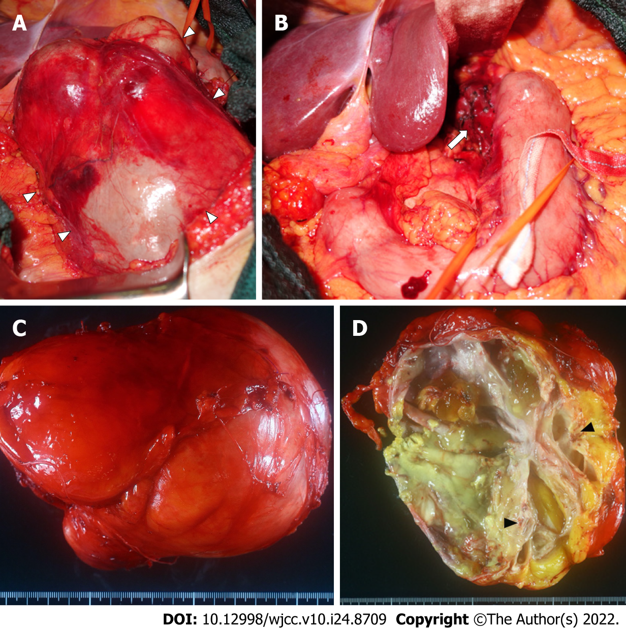
Figure 3 Intraoperative findings and macroscopic findings of the resected specimen.
A: A smooth surfaced mass (white arrowhead) is exposed after resection of the lesser omentum; B: The mass is resected with a part of the seromuscular layer of the lessor curvature of the stomach (white arrow) and removed; C: The resected specimen is a multifocal mass filled with viscous mucus, 15 cm × 12 cm × 12 cm in size and weighing 1240 g; D: Cartilage-like tissue (black arrowhead) is observed in part of the cystic wall.
- Citation: Murakami T, Shimizu H, Yamazaki K, Nojima H, Usui A, Kosugi C, Shuto K, Obi S, Sato T, Yamazaki M, Koda K. Intra-abdominal ectopic bronchogenic cyst with a mucinous neoplasm harboring a GNAS mutation: A case report. World J Clin Cases 2022; 10(24): 8709-8717
- URL: https://www.wjgnet.com/2307-8960/full/v10/i24/8709.htm
- DOI: https://dx.doi.org/10.12998/wjcc.v10.i24.8709


