Copyright
©The Author(s) 2022.
World J Clin Cases. Aug 26, 2022; 10(24): 8695-8702
Published online Aug 26, 2022. doi: 10.12998/wjcc.v10.i24.8695
Published online Aug 26, 2022. doi: 10.12998/wjcc.v10.i24.8695
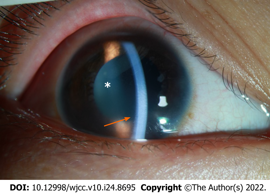
Figure 1 Image of the anterior chamber in the right eye on Day 1 after hospitalization.
The arrow shows corneal edema with haze. The depth of the anterior chamber was normal. Mutton-fat KP: ++ (arrow); anterior chamber cells: +++ (asterisk).
- Citation: Zhang Y, Tang L. Retinoblastoma in an older child with secondary glaucoma as the first clinical presenting symptom: A case report. World J Clin Cases 2022; 10(24): 8695-8702
- URL: https://www.wjgnet.com/2307-8960/full/v10/i24/8695.htm
- DOI: https://dx.doi.org/10.12998/wjcc.v10.i24.8695


