Copyright
©The Author(s) 2022.
World J Clin Cases. Aug 16, 2022; 10(23): 8406-8416
Published online Aug 16, 2022. doi: 10.12998/wjcc.v10.i23.8406
Published online Aug 16, 2022. doi: 10.12998/wjcc.v10.i23.8406
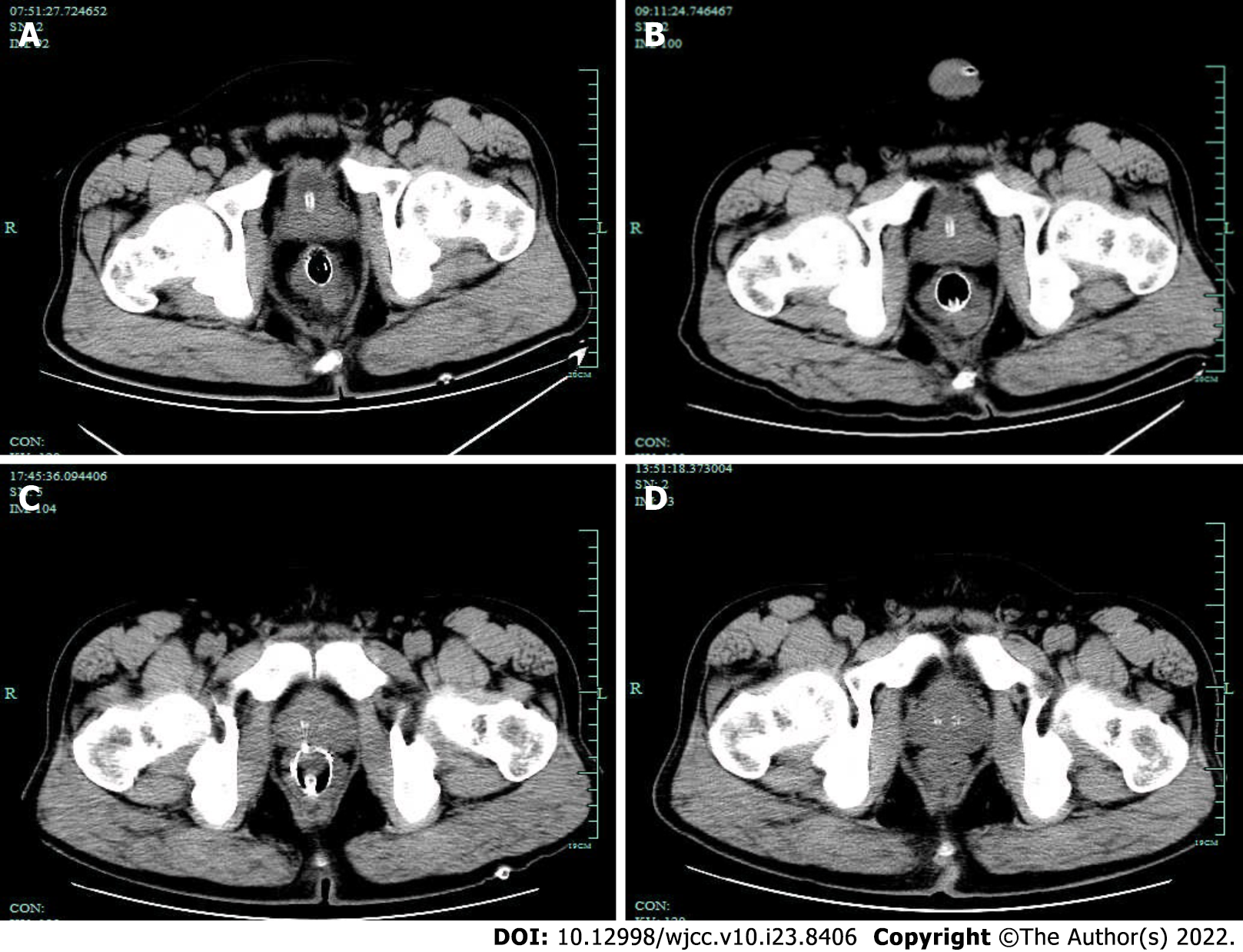
Figure 7 The pelvic free gas gradually decreased in the patient.
A: Day 2 computed tomography (CT), Metal tubular shadows were observed in the rectal lumen, and a lot of gas was observed around the intestinal canal; B: Day 5 CT: a lot of free air was trapped behind the peritoneum and in the pelvis, but this was slightly resolved on day 5, as compared the amount of air on Day 2 CT slice; C: Day 10 CT: The free air in the peritoneal cavity and behind the peritoneum was significantly reduced as compared with that on Day 5 CT slice; D: Day 17 CT: No clear manifestation of the original rectal stent was observed. A little amount of free air trapped in the pelvis was absorbed as compared with that on the Day 10 CT slice.
- Citation: Cheng SL, Xie L, Wu HW, Zhang XF, Lou LL, Shen HZ. Metal stent combined with ileus drainage tube for the treatment of delayed rectal perforation: A case report. World J Clin Cases 2022; 10(23): 8406-8416
- URL: https://www.wjgnet.com/2307-8960/full/v10/i23/8406.htm
- DOI: https://dx.doi.org/10.12998/wjcc.v10.i23.8406


