Copyright
©The Author(s) 2022.
World J Clin Cases. Aug 16, 2022; 10(23): 8392-8399
Published online Aug 16, 2022. doi: 10.12998/wjcc.v10.i23.8392
Published online Aug 16, 2022. doi: 10.12998/wjcc.v10.i23.8392
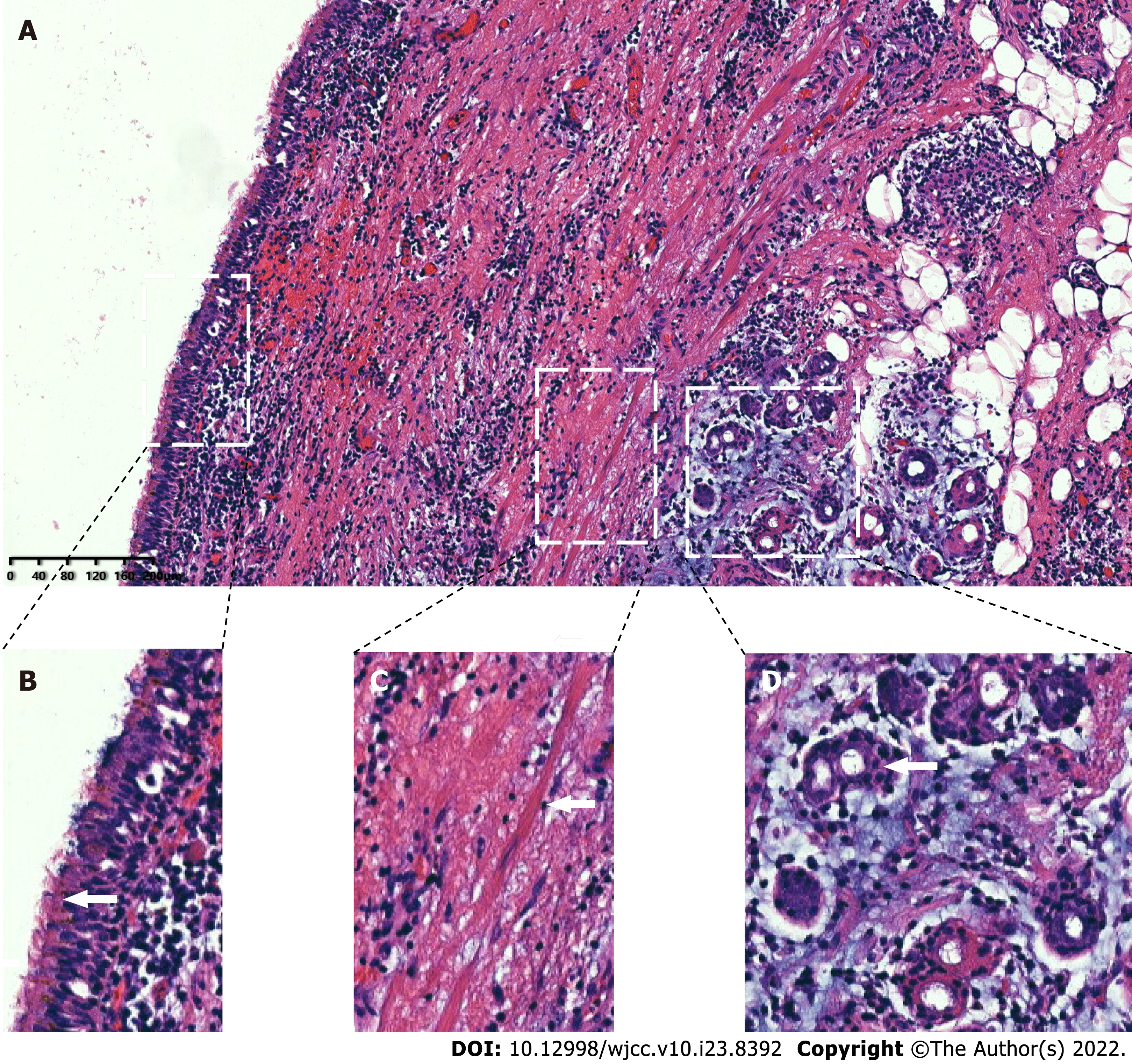
Figure 3 Postoperative histopathology.
A: Combined with immunohistochemical staining for CK7 (+), CK20 (-), TTF1 (-) and S-100 (-), the histopathological examinations showed that the specimen was consistent with a bronchogenic cyst with obvious hyperplasia of histiocytes (× 100); B: The white arrows represent column-like cilia epithelial cells; C: Smooth muscle bundles; D: Bronchial glands.
- Citation: Ma B, Fu KW, Xie XD, Cheng Y, Wang SQ. Bronchogenic cysts with infection in the chest wall skin of a 64-year-old asymptomatic patient: A case report. World J Clin Cases 2022; 10(23): 8392-8399
- URL: https://www.wjgnet.com/2307-8960/full/v10/i23/8392.htm
- DOI: https://dx.doi.org/10.12998/wjcc.v10.i23.8392


