Copyright
©The Author(s) 2022.
World J Clin Cases. Aug 16, 2022; 10(23): 8375-8383
Published online Aug 16, 2022. doi: 10.12998/wjcc.v10.i23.8375
Published online Aug 16, 2022. doi: 10.12998/wjcc.v10.i23.8375
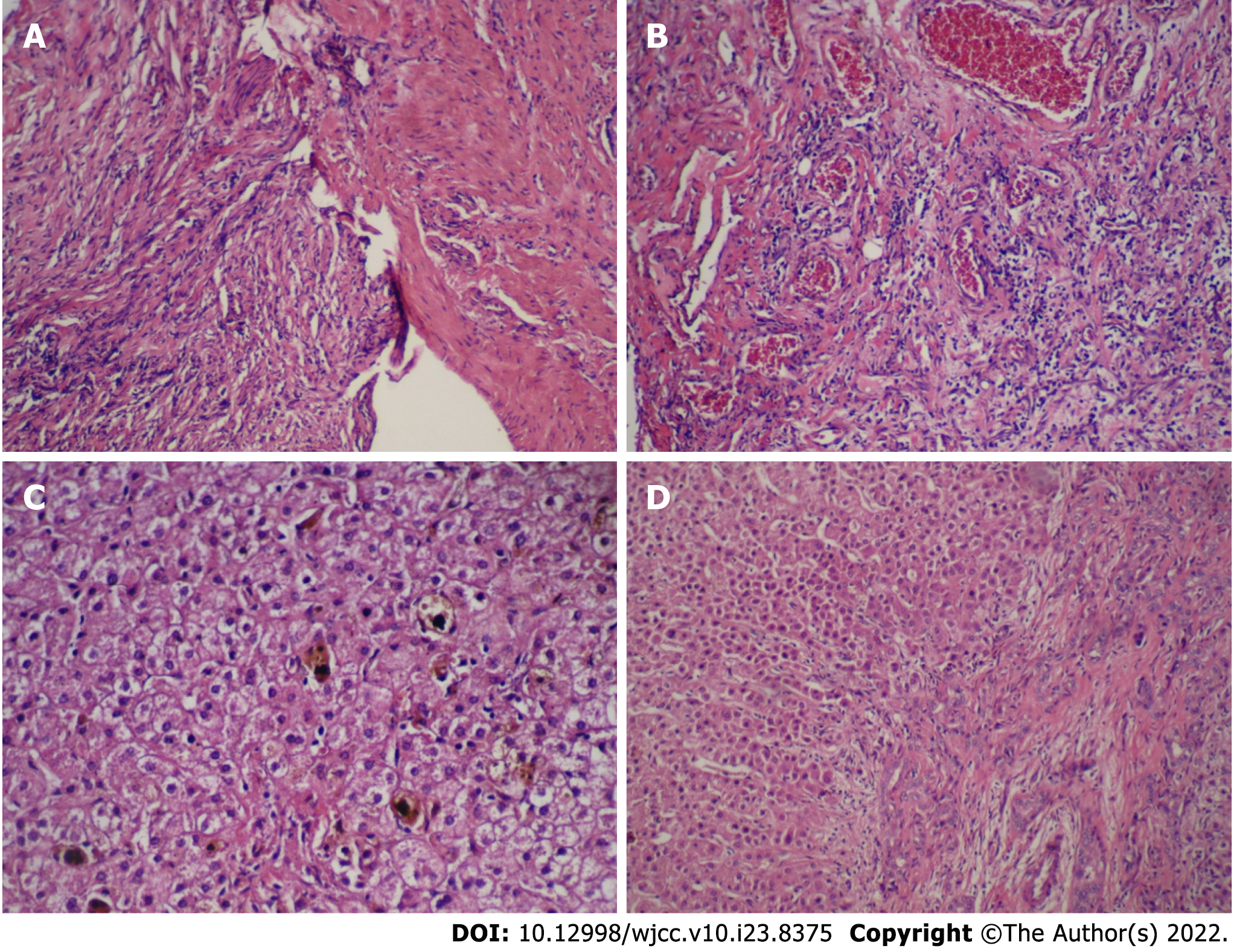
Figure 2 The hematoxylin-eosin staining for the tumor and the liver tissue.
A and B: Inflammatory myofibroblastic tumor of the biliary duct composed of spindle cells, fibrous tissue and abundant small vessels in a background of inflammatory cellular infiltration and myxoid stroma (A: Original magnification: 100 ×; scale bar: 100 μm; and B: Original magnification: 400 ×; scale bar: 100 μm.); C and D: The bile canaliculus hyperplasia, hepatic fibrosis, and lymphocytes infiltration accompanied with hepatocyte degeneration and cholestasis could be observed at portal area (C: Original magnification: 400 ×; scale bar: 100 μm; and D: Original magnification: 100 ×; scale bar: 100 μm).
- Citation: Huang Y, Shu SN, Zhou H, Liu LL, Fang F. Infant biliary cirrhosis secondary to a biliary inflammatory myofibroblastic tumor: A case report and review of literature. World J Clin Cases 2022; 10(23): 8375-8383
- URL: https://www.wjgnet.com/2307-8960/full/v10/i23/8375.htm
- DOI: https://dx.doi.org/10.12998/wjcc.v10.i23.8375


