Copyright
©The Author(s) 2022.
World J Clin Cases. Aug 16, 2022; 10(23): 8367-8374
Published online Aug 16, 2022. doi: 10.12998/wjcc.v10.i23.8367
Published online Aug 16, 2022. doi: 10.12998/wjcc.v10.i23.8367
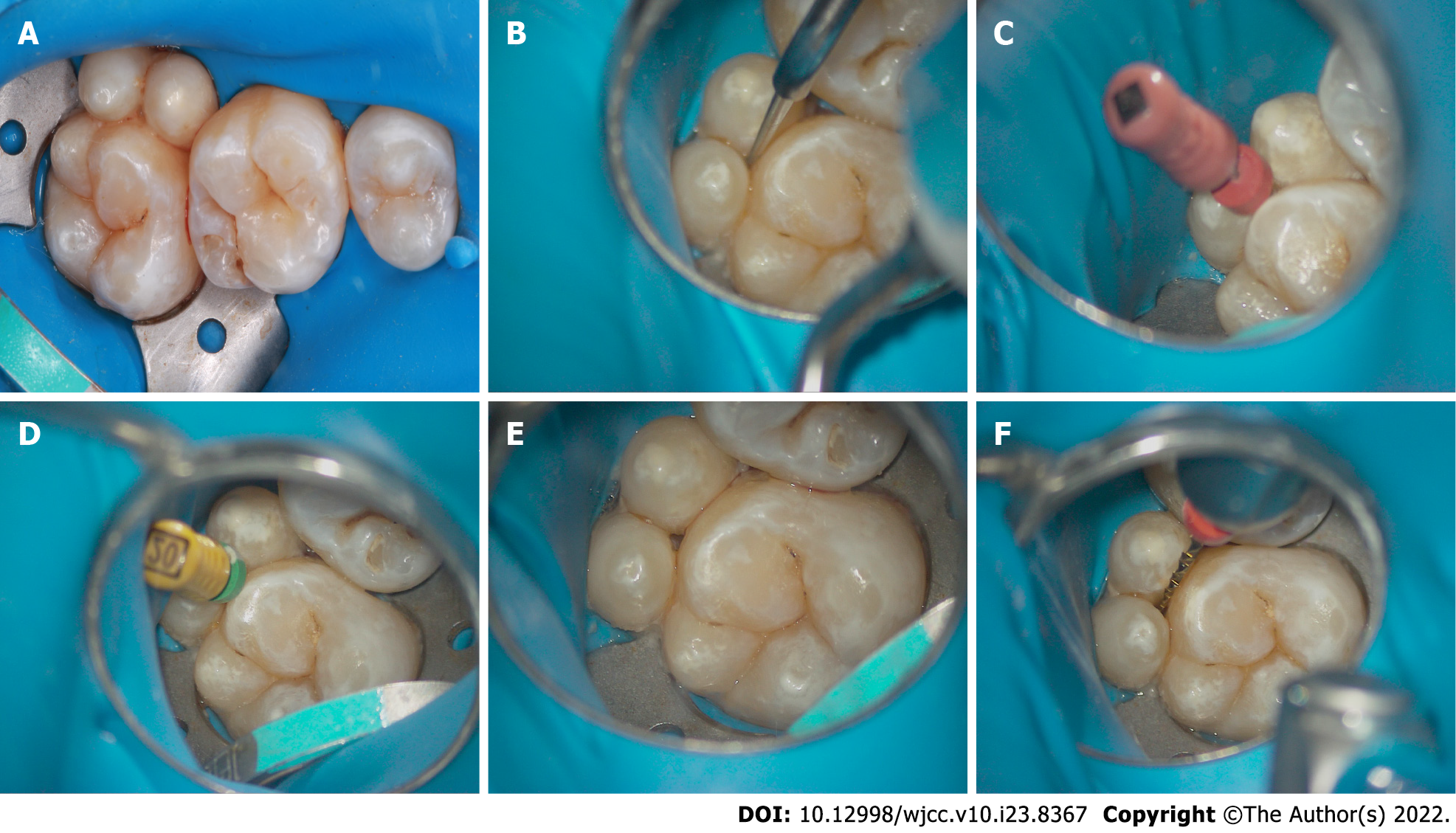
Figure 2 Representative intraoral photos of the fused tooth during root canal therapy.
A: A rubber dam was positioned; B: A ball drill (ET BD) and ET 20 of the ultrasonic equipment (P5 Newtron) were used to remove the damaged tissue under a microscope; C: An electronic apex locator was utilized to determine the working lengths with 6#K-file; D-E: Filing was performed until size 20# was reached; F: Waveone Gold Ni-Ti rotary instruments were used to clean and shape the canals.
- Citation: Mei XH, Liu J, Wang W, Zhang QX, Hong T, Bai SZ, Cheng XG, Tian Y, Jiang WK. Endodontic management of a fused left maxillary second molar and two paramolars using cone beam computed tomography: A case report. World J Clin Cases 2022; 10(23): 8367-8374
- URL: https://www.wjgnet.com/2307-8960/full/v10/i23/8367.htm
- DOI: https://dx.doi.org/10.12998/wjcc.v10.i23.8367


