Copyright
©The Author(s) 2022.
World J Clin Cases. Aug 16, 2022; 10(23): 8367-8374
Published online Aug 16, 2022. doi: 10.12998/wjcc.v10.i23.8367
Published online Aug 16, 2022. doi: 10.12998/wjcc.v10.i23.8367
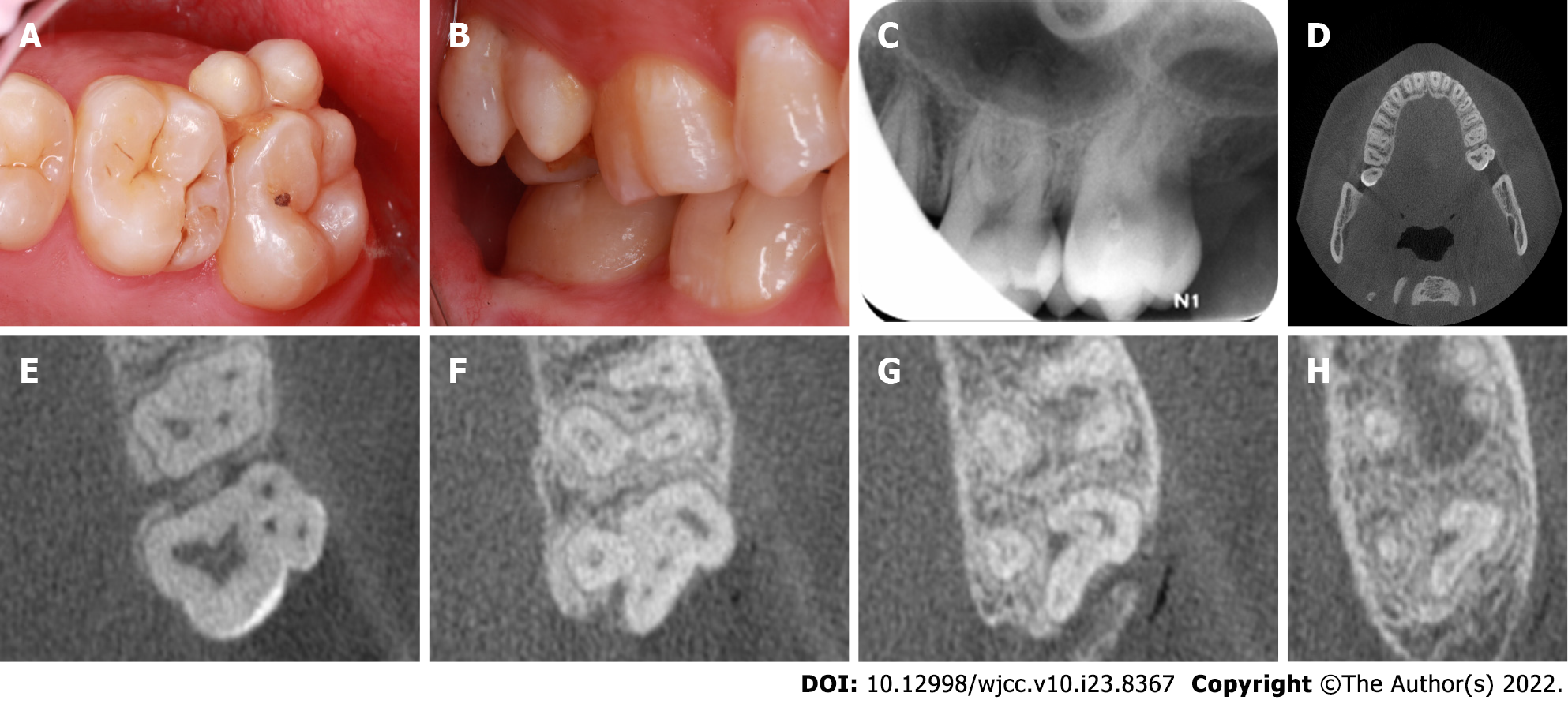
Figure 1 Representative preoperative intraoral photographs and radiographs of the fusion tooth.
A: Occlusal intraoral view revealing the presence of two supernumerary paramolars on the buccal aspect of the maxillary left second molar, with evidence of distinct developmental grooves separating these teeth and additional evidence of food impaction; B: Buccal view; C: A preoperative radiograph of the left maxillary second molar; D: Cone-beam computed tomography (CBCT) axial cross-sections of the maxillary fused second molar and paramolars; E-H: CBCT images revealed three slightly curved, patent root canals, which were associated with the maxillary second molar, and there was single similar canal for each of the fused paramolars. The apex of the irregular bending isthmus area that was formed by the intersection of three teeth was connected in a periapical transmission image.
- Citation: Mei XH, Liu J, Wang W, Zhang QX, Hong T, Bai SZ, Cheng XG, Tian Y, Jiang WK. Endodontic management of a fused left maxillary second molar and two paramolars using cone beam computed tomography: A case report. World J Clin Cases 2022; 10(23): 8367-8374
- URL: https://www.wjgnet.com/2307-8960/full/v10/i23/8367.htm
- DOI: https://dx.doi.org/10.12998/wjcc.v10.i23.8367


