Copyright
©The Author(s) 2022.
World J Clin Cases. Aug 16, 2022; 10(23): 8344-8351
Published online Aug 16, 2022. doi: 10.12998/wjcc.v10.i23.8344
Published online Aug 16, 2022. doi: 10.12998/wjcc.v10.i23.8344
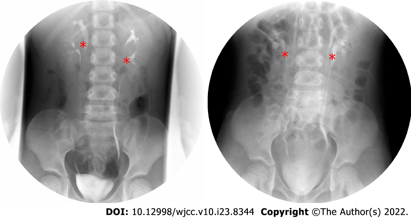
Figure 1 Intravenous urography.
The left side image was taken before operation. The right side image was taken three months after the operation. The double renal pelvis and double ureter malformations were identified by asterisk, and bilateral ureters were unobstructed and no hydronephrosis was found after operation.
- Citation: Wang SB, Wan L, Wang Y, Yi ZJ, Xiao C, Cao JZ, Liu XY, Tang RP, Luo Y. Laparoscopic treatment of bilateral duplex kidney and ectopic ureter: A case report. World J Clin Cases 2022; 10(23): 8344-8351
- URL: https://www.wjgnet.com/2307-8960/full/v10/i23/8344.htm
- DOI: https://dx.doi.org/10.12998/wjcc.v10.i23.8344


