Copyright
©The Author(s) 2022.
World J Clin Cases. Aug 16, 2022; 10(23): 8304-8311
Published online Aug 16, 2022. doi: 10.12998/wjcc.v10.i23.8304
Published online Aug 16, 2022. doi: 10.12998/wjcc.v10.i23.8304
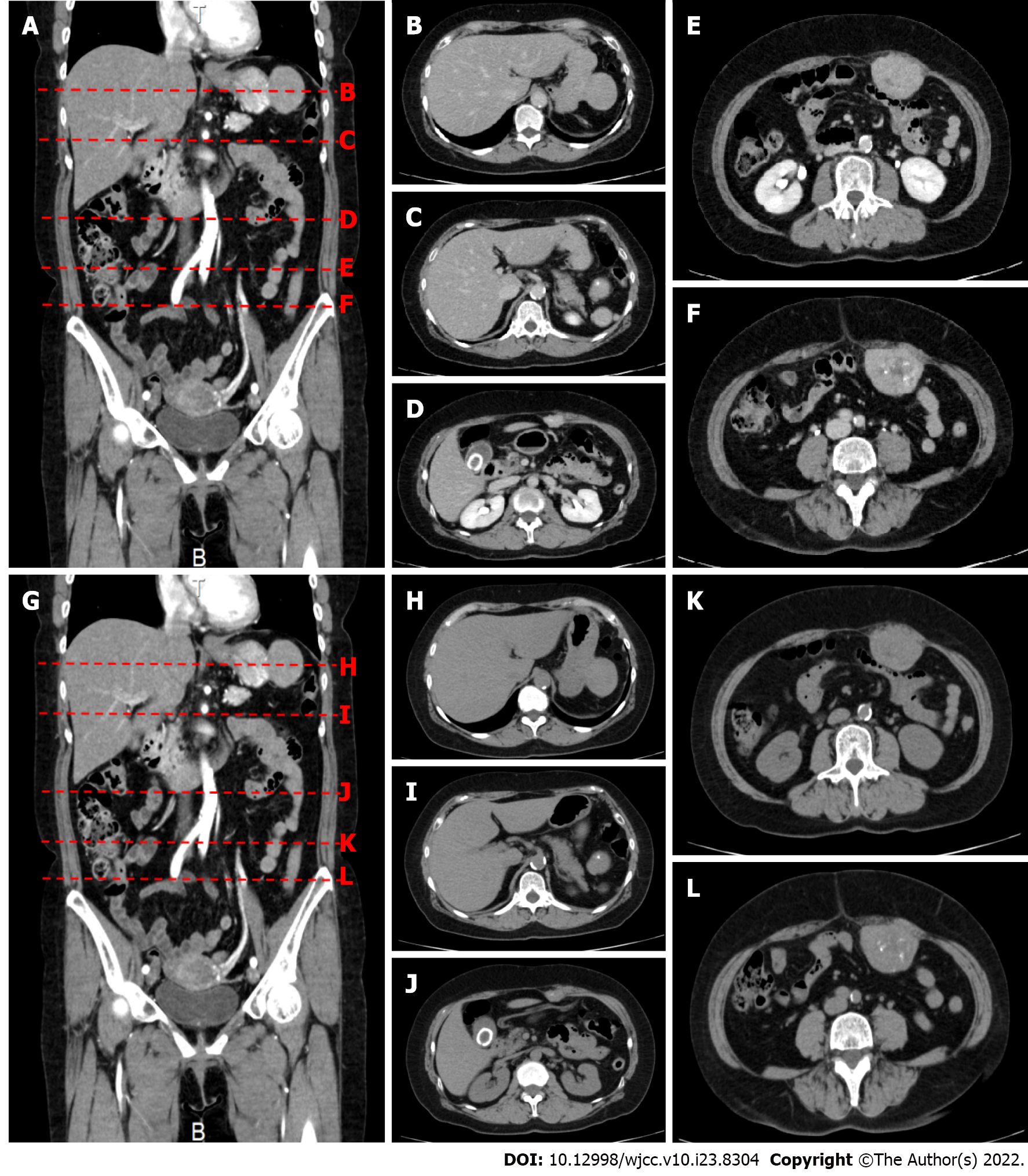
Figure 1 Contrast-enhanced computed tomography shows a 50 mm × 40 mm × 48 mm mass protruding from below the serosa at the greater curvature of the upper stomach.
A: Contrast enhancement is observed in the arterial phase; B and H: A 50 mm × 40 mm × 48 mm mass protrudes outward from the greater curvature of the upper stomach; C and I: A nodule about 7 mm in size is found near the accessory spleen; D and J: Three masses protruding from the abdominal wall into the peritoneal cavity are also observed. The most cranial abdominal wall mass measures 28 mm × 16 mm × 18 mm and has irregular margins; E and K: The middle abdominal wall mass measures 55 mm × 42 mm × 36 mm and has irregular margins; F and L: The most caudal abdominal wall mass measures 55 mm × 44 mm × 55 mm and has irregular margins. All of these masses show internal calcification and contrast enhancement; G: Plain computed tomography.
- Citation: Ohkura Y, Uruga H, Shiiba M, Ito S, Shimoyama H, Ishihara M, Ueno M, Udagawa H. Phosphoglyceride crystal deposition disease requiring differential diagnosis from malignant tumors and confirmed by Raman spectroscopy: A case report. World J Clin Cases 2022; 10(23): 8304-8311
- URL: https://www.wjgnet.com/2307-8960/full/v10/i23/8304.htm
- DOI: https://dx.doi.org/10.12998/wjcc.v10.i23.8304


