Copyright
©The Author(s) 2022.
World J Clin Cases. Aug 16, 2022; 10(23): 8291-8297
Published online Aug 16, 2022. doi: 10.12998/wjcc.v10.i23.8291
Published online Aug 16, 2022. doi: 10.12998/wjcc.v10.i23.8291
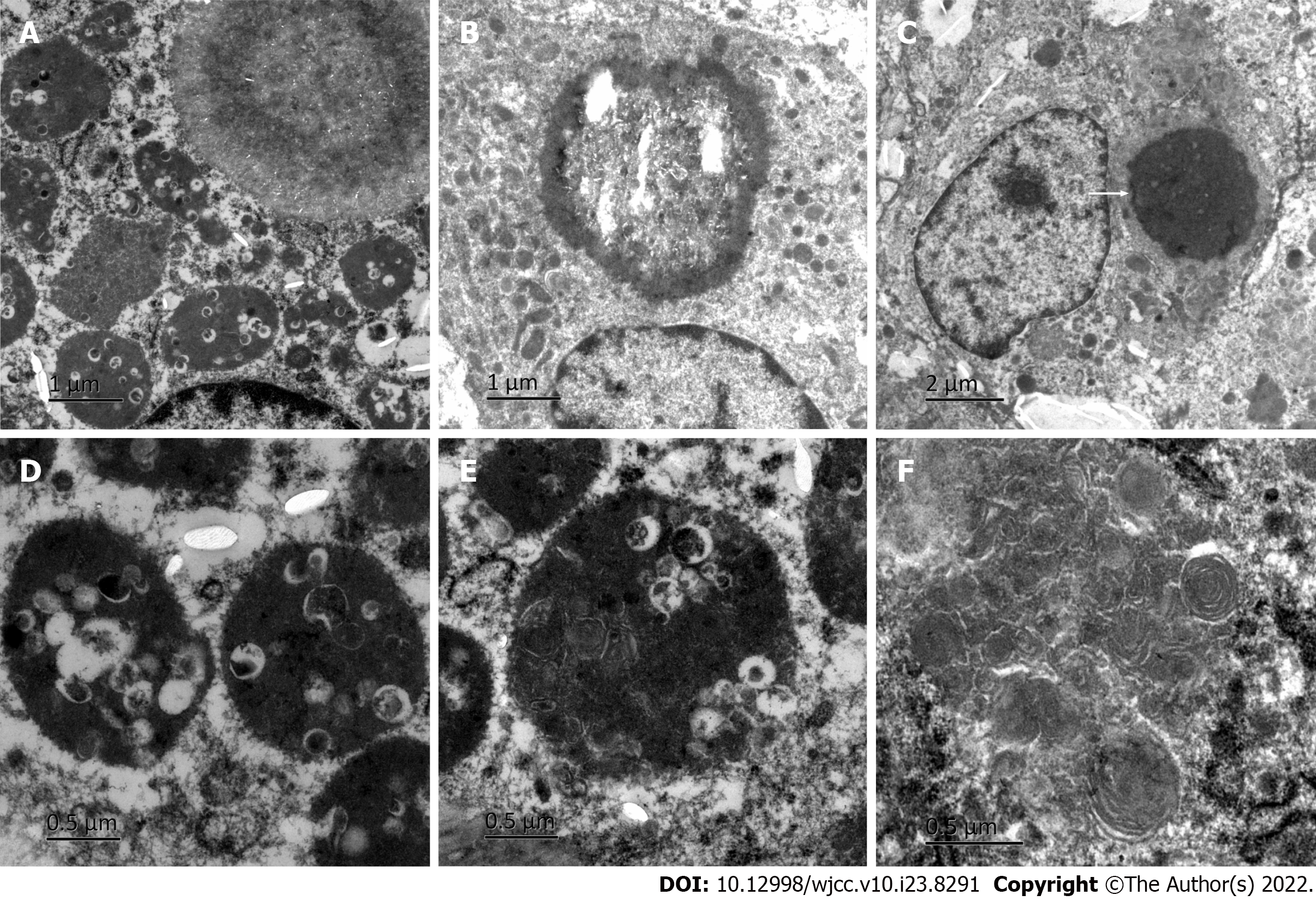
Figure 4 Electron microscopy findings.
A: Phagocytic lysosomes in the cytoplasm of macrophages; B: Microbubble; C: A typical Michaelis–Gutman (M-G) body as shown by arrows; D: Large number of bacteria in phagocytic lysosomes; E: Some bacteria were digested and dissolved; F: Myelin figure.
- Citation: Wang HK, Hang G, Wang YY, Wen Q, Chen B. Bladder malacoplakia: A case report. World J Clin Cases 2022; 10(23): 8291-8297
- URL: https://www.wjgnet.com/2307-8960/full/v10/i23/8291.htm
- DOI: https://dx.doi.org/10.12998/wjcc.v10.i23.8291


