Copyright
©The Author(s) 2022.
World J Clin Cases. Aug 16, 2022; 10(23): 8277-8283
Published online Aug 16, 2022. doi: 10.12998/wjcc.v10.i23.8277
Published online Aug 16, 2022. doi: 10.12998/wjcc.v10.i23.8277
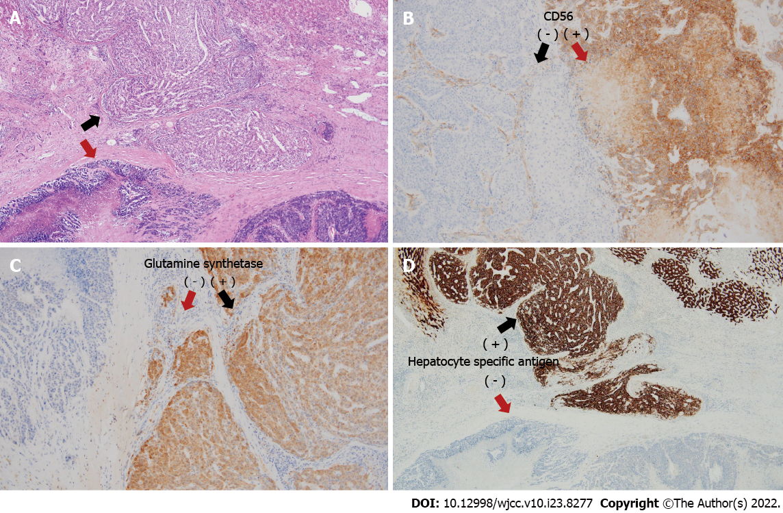
Figure 4 Histopathological analysis and immunohistochemical examination of the resected specimen.
The collision tumor comprises two distinct components: large-cell neuroendocrine carcinoma (red arrow) and hepatocellular carcinoma (black arrow). A: Hematoxylin-eosin staining (× 40); B and C: Immunohistochemical staining (B) for CD56 (× 100) and (C) glutamine synthetase staining (× 100); D: Hepatocyte-specific antigen staining (× 40).
- Citation: Noh BG, Seo HI, Park YM, Kim S, Hong SB, Lee SJ. Complete resection of large-cell neuroendocrine and hepatocellular carcinoma of the liver: A case report. World J Clin Cases 2022; 10(23): 8277-8283
- URL: https://www.wjgnet.com/2307-8960/full/v10/i23/8277.htm
- DOI: https://dx.doi.org/10.12998/wjcc.v10.i23.8277


