Copyright
©The Author(s) 2022.
World J Clin Cases. Aug 16, 2022; 10(23): 8232-8241
Published online Aug 16, 2022. doi: 10.12998/wjcc.v10.i23.8232
Published online Aug 16, 2022. doi: 10.12998/wjcc.v10.i23.8232
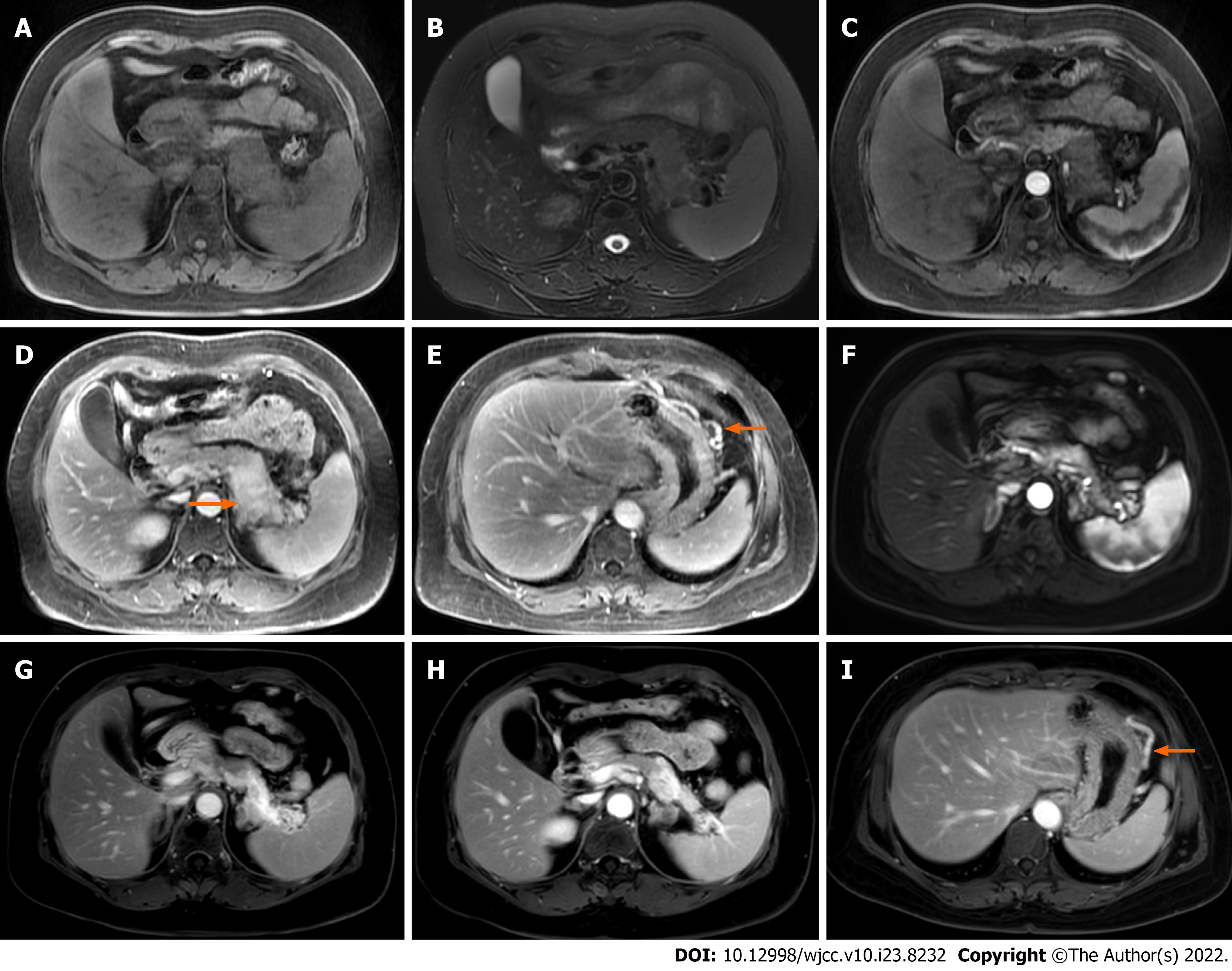
Figure 2 Focal autoimmune pancreatitis with progressive fibrosis after spontaneous remission in case 2.
A and B: Enlargement of the pancreatic body and tail; C, D, and E: In addition to the gradual enhancement of the lesion in the body and tail of the pancreas, stenosis of the splenic vein (arrow) and dilation of the large curved side vein of the gastric body (arrow) are observed; F and G: Magnetic resonance imaging (MRI) follow-up at 22 mo showing that the body and tail of the pancreas is significantly reduced and more obviously gradually enhanced; H: Lower level MRI showing that the volume of the pancreatic body and tail is significantly atrophied and obviously enhanced; I: The dilation of the large curvature side vein of the gastric body (arrow) is not improved.
- Citation: Zhang BB, Huo JW, Yang ZH, Wang ZC, Jin EH. Spontaneous remission of autoimmune pancreatitis: Four case reports . World J Clin Cases 2022; 10(23): 8232-8241
- URL: https://www.wjgnet.com/2307-8960/full/v10/i23/8232.htm
- DOI: https://dx.doi.org/10.12998/wjcc.v10.i23.8232


