Copyright
©The Author(s) 2022.
World J Clin Cases. Aug 16, 2022; 10(23): 8232-8241
Published online Aug 16, 2022. doi: 10.12998/wjcc.v10.i23.8232
Published online Aug 16, 2022. doi: 10.12998/wjcc.v10.i23.8232
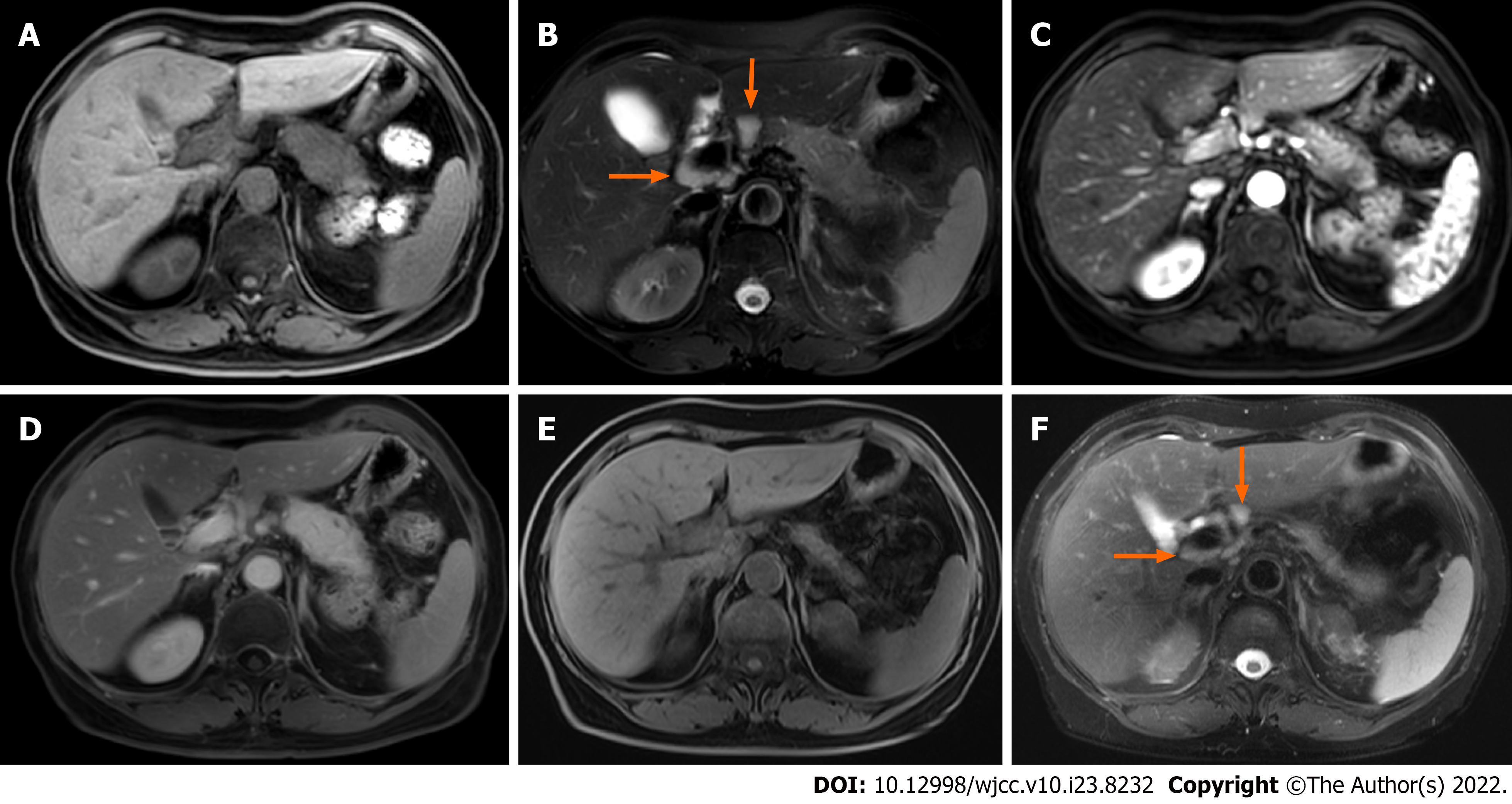
Figure 1 Focal autoimmune pancreatitis with atrophy after spontaneous remission in case 1.
A and B: The pancreatic body and tail enlarge, and multiple enlarged lymph nodes (arrows) are seen around the portal vein; C and D: Gradual enhancement of the lesion on enhanced scan; E and F: Magnetic resonance imaging follow-up at the 13th month. Pancreatic swelling dissipates and the volume of the pancreas decreases significantly, and the enlarged lymph nodes (arrows) are significantly reduced as well.
- Citation: Zhang BB, Huo JW, Yang ZH, Wang ZC, Jin EH. Spontaneous remission of autoimmune pancreatitis: Four case reports . World J Clin Cases 2022; 10(23): 8232-8241
- URL: https://www.wjgnet.com/2307-8960/full/v10/i23/8232.htm
- DOI: https://dx.doi.org/10.12998/wjcc.v10.i23.8232


