Copyright
©The Author(s) 2022.
World J Clin Cases. Aug 16, 2022; 10(23): 8063-8075
Published online Aug 16, 2022. doi: 10.12998/wjcc.v10.i23.8063
Published online Aug 16, 2022. doi: 10.12998/wjcc.v10.i23.8063
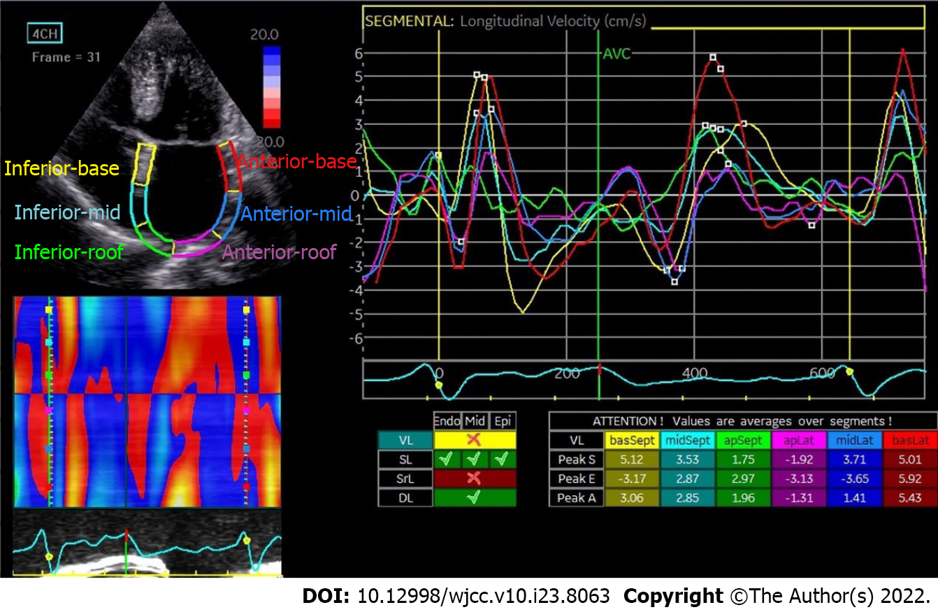
Figure 3 Atrial strain image by 2D-speckle tracking echocardiography in a patient with atrial fibrillation.
Similarly, as shown on the left, left atrium is divided into six segments distinguished by color and corresponds to the strain curve in the same color from the apical four-chamber view. Each segment of left atrial diastole and contraction successively over time is shown on the right. The strain curves in patients with atrial fibrillation (AF) are more disorganized than those in healthy volunteers (Figure 1). The different stages of atrial function cannot be clearly distinguished over the cardiac cycle in AF patients.
- Citation: Yu ZX, Yang W, Yin WS, Peng KX, Pan YL, Chen WW, Du BB, He YQ, Yang P. Clinical utility of left atrial strain in predicting atrial fibrillation recurrence after catheter ablation: An up-to-date review. World J Clin Cases 2022; 10(23): 8063-8075
- URL: https://www.wjgnet.com/2307-8960/full/v10/i23/8063.htm
- DOI: https://dx.doi.org/10.12998/wjcc.v10.i23.8063


