Copyright
©The Author(s) 2022.
World J Clin Cases. Aug 6, 2022; 10(22): 7960-7967
Published online Aug 6, 2022. doi: 10.12998/wjcc.v10.i22.7960
Published online Aug 6, 2022. doi: 10.12998/wjcc.v10.i22.7960
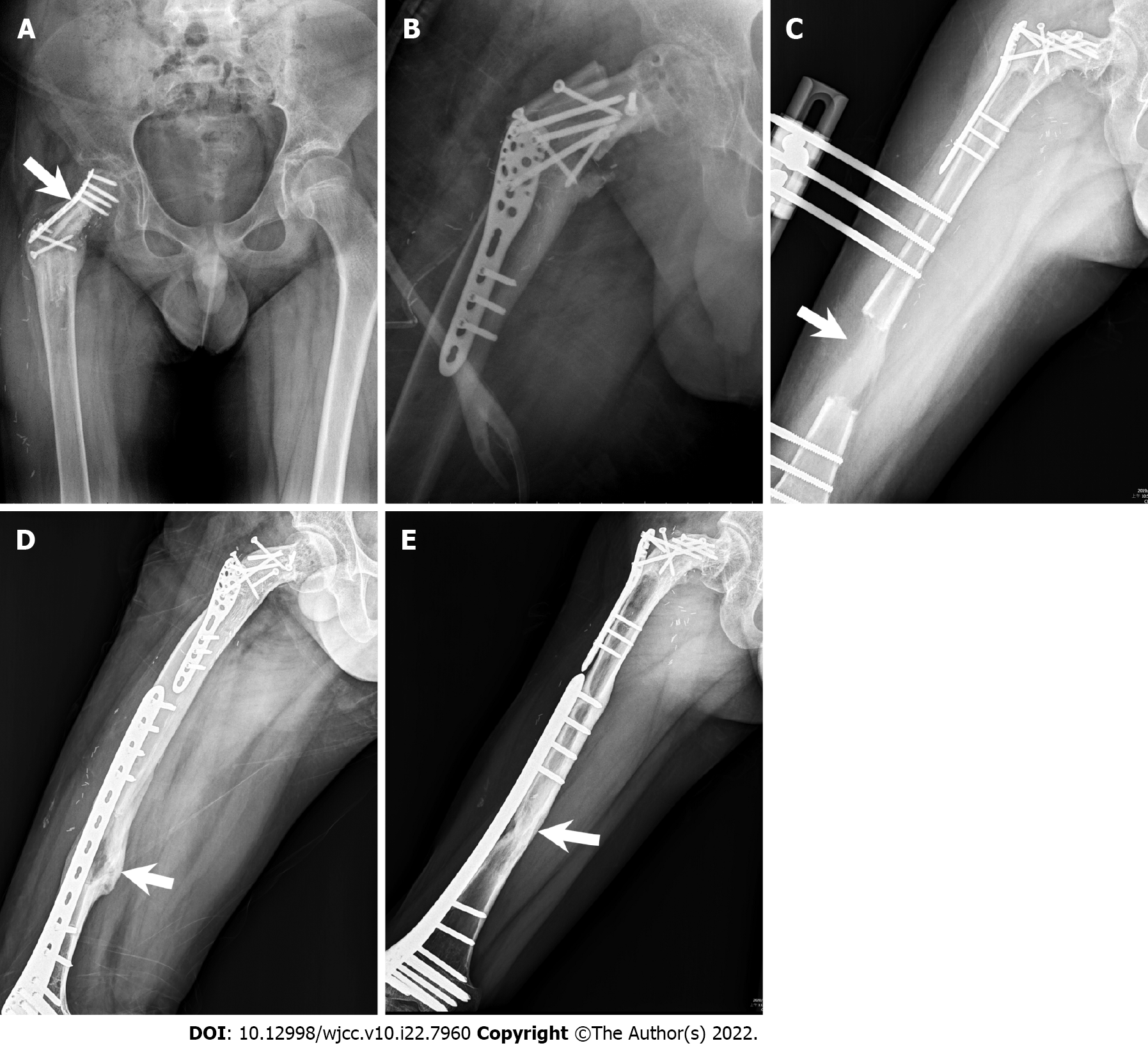
Figure 5 Radiographic images.
A: Plain X-ray film obtained after an increase in partial weight bearing. The plate was deformed (arrow: bending site of the plate); B: Revision and fixation with Arbeitsgemeinschaft für Osteosynthesefragen proximal humeral internal locking system plate; C: Radiographic image of orthosis with complete distraction. The femoral distraction gap is approximately 6.5 cm (arrow: distraction gap); D: Radiographic image showing removal of the orthosis and international fixation with less-invasive stabilization system complete corticalization (arrow: corticalized bone); E: Radiographic image showing complete consolidation phase (arrow: cordialized bone with increased radiopacity).
- Citation: Lai CY, Chen KJ, Ho TY, Li LY, Kuo CC, Chen HT, Fong YC. Resection with limb salvage in an Asian male adolescent with Ewing’s sarcoma: A case report. World J Clin Cases 2022; 10(22): 7960-7967
- URL: https://www.wjgnet.com/2307-8960/full/v10/i22/7960.htm
- DOI: https://dx.doi.org/10.12998/wjcc.v10.i22.7960


