Copyright
©The Author(s) 2022.
World J Clin Cases. Aug 6, 2022; 10(22): 7960-7967
Published online Aug 6, 2022. doi: 10.12998/wjcc.v10.i22.7960
Published online Aug 6, 2022. doi: 10.12998/wjcc.v10.i22.7960
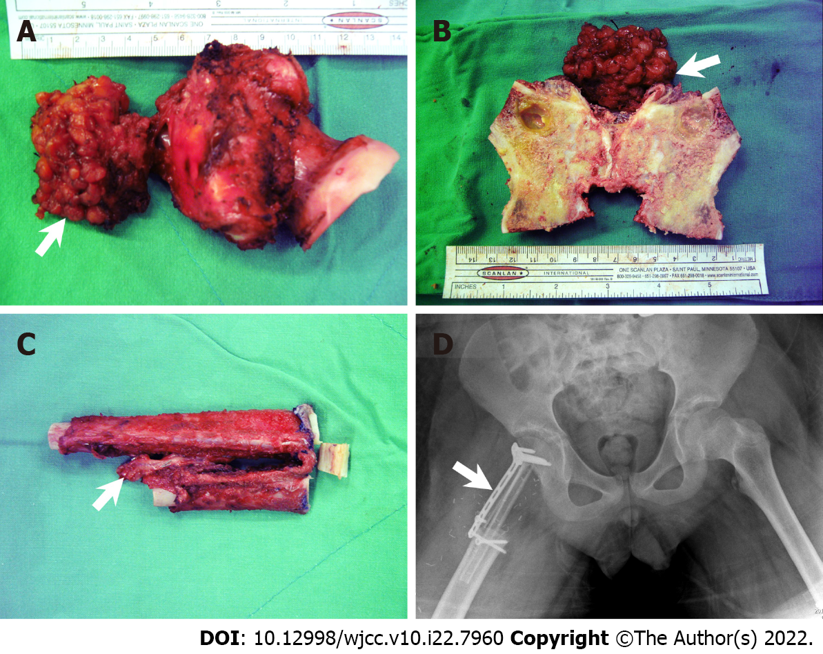
Figure 4 Intraoperative sample and postoperative pelvic plain imaging.
A: Gross view of the resected tumor and resected femoral neck (arrow: Ewing’s sarcoma extra-bony part); B: Split resected femoral neck; C: Folded autologous fibular graft (arrow: vascular pedicle); D: Pelvic plain imaging (anteroposterior) obtained immediately after the surgery (arrow: locking plate used to fix the fibular graft).
- Citation: Lai CY, Chen KJ, Ho TY, Li LY, Kuo CC, Chen HT, Fong YC. Resection with limb salvage in an Asian male adolescent with Ewing’s sarcoma: A case report. World J Clin Cases 2022; 10(22): 7960-7967
- URL: https://www.wjgnet.com/2307-8960/full/v10/i22/7960.htm
- DOI: https://dx.doi.org/10.12998/wjcc.v10.i22.7960


