Copyright
©The Author(s) 2022.
World J Clin Cases. Aug 6, 2022; 10(22): 7944-7949
Published online Aug 6, 2022. doi: 10.12998/wjcc.v10.i22.7944
Published online Aug 6, 2022. doi: 10.12998/wjcc.v10.i22.7944
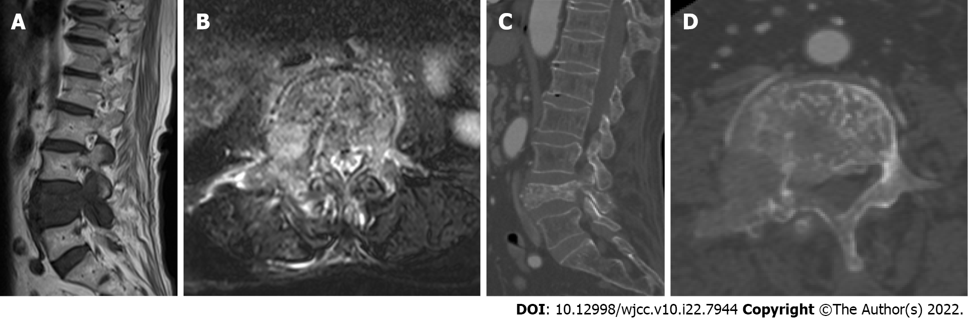
Figure 1 Computed tomography and magnetic resonance imaging.
A: Sagittal position lumbar magnetic resonance imaging (MRI) showed a huge metastatic mass destroying L4 vertebral body and pedicle and compressing the nerve root; B: Cross-sectional lumbar MRI showed vertebral body and pedicle were damaged; C: L4 vertebral body and pedicle pathological fractured; D: L4 vertebral body and pedicle were damaged.
- Citation: Ran Q, Li T, Kuang ZP, Guo XH. Percutaneous transforaminal endoscopic decompression combined with percutaneous vertebroplasty in treatment of lumbar vertebral body metastases: A case report. World J Clin Cases 2022; 10(22): 7944-7949
- URL: https://www.wjgnet.com/2307-8960/full/v10/i22/7944.htm
- DOI: https://dx.doi.org/10.12998/wjcc.v10.i22.7944


