Copyright
©The Author(s) 2022.
World J Clin Cases. Aug 6, 2022; 10(22): 7913-7923
Published online Aug 6, 2022. doi: 10.12998/wjcc.v10.i22.7913
Published online Aug 6, 2022. doi: 10.12998/wjcc.v10.i22.7913
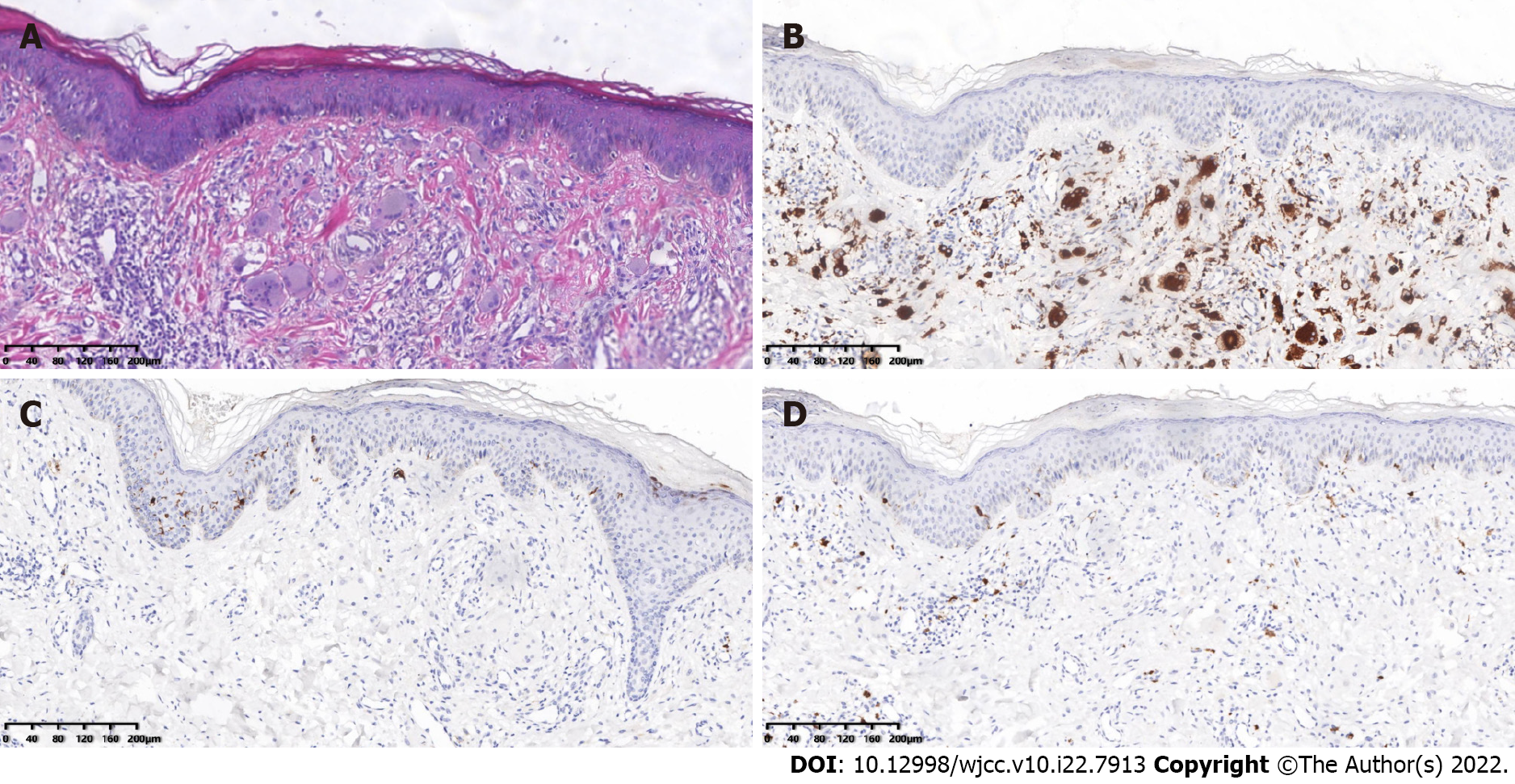
Figure 3 Pathological examination of the patient’s skin.
A: Histopathology of the skin from the left upper back. The epidermis was generally normal, and the dermal papillae showed an increased number of histiocytes and multinucleated giant cells with abundant cell cytoplasm and hairy glass-like changes, with a little surrounding lymphatic and eosinophilic infiltration (HE 40×). Immunohistochemical staining: B: CD68-positive; C: CD1a negative; and D: S-100 negative (SP×100).
- Citation: Xu XL, Liang XH, Liu J, Deng X, Zhang L, Wang ZG. Multicentric reticulohistiocytosis with prominent skin lesions and arthritis: A case report. World J Clin Cases 2022; 10(22): 7913-7923
- URL: https://www.wjgnet.com/2307-8960/full/v10/i22/7913.htm
- DOI: https://dx.doi.org/10.12998/wjcc.v10.i22.7913


