Copyright
©The Author(s) 2022.
World J Clin Cases. Aug 6, 2022; 10(22): 7883-7889
Published online Aug 6, 2022. doi: 10.12998/wjcc.v10.i22.7883
Published online Aug 6, 2022. doi: 10.12998/wjcc.v10.i22.7883
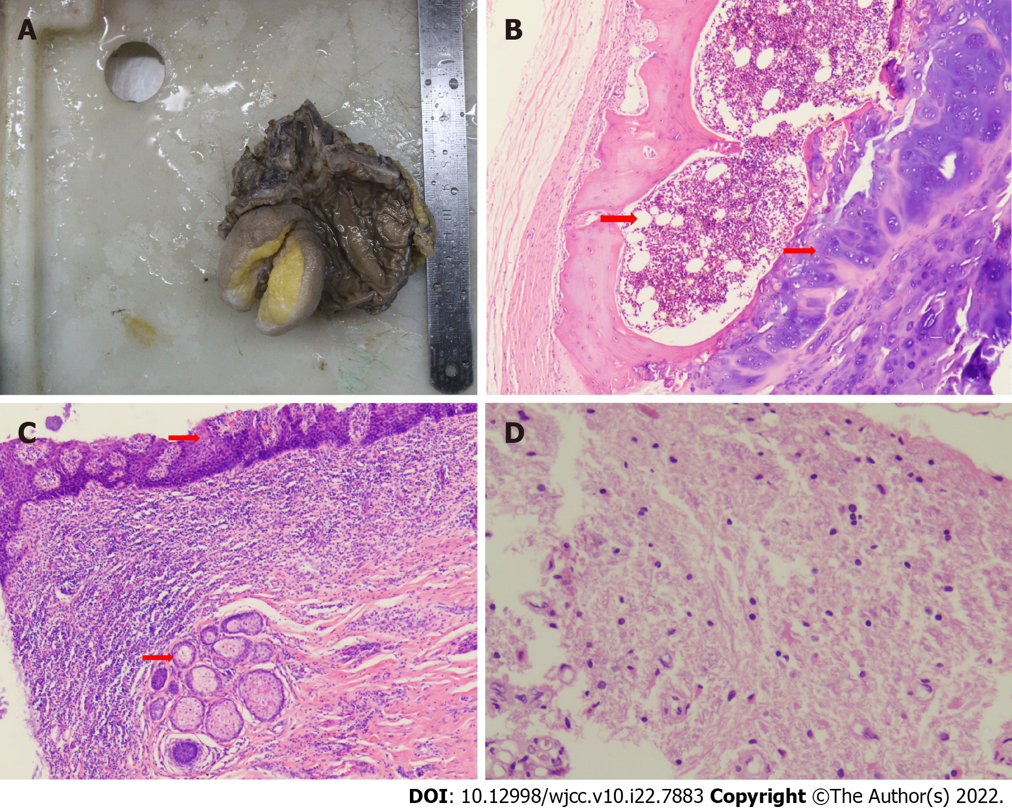
Figure 4 Pathology of rectal teratoma.
A: Surgical resection of pathological specimens. The mass was completely removed, and a piece was removed every 1 cm. The tissues were then fixed, dehydrated, soaked in wax, embedded, and stained with hematoxylin and eosin staining (HE); B: Rectal teratoma with bone marrow and cartilage (indicated by the arrow) (HE, 4 × 10 magnification); C: Rectal teratoma with squamous epithelium and sebaceous glands (indicated by the arrow) (HE, 4 × 10 magnification); D: Rectal teratoma with brain tissue (HE, 20 × 10 magnification).
- Citation: Liu JL, Sun PL. Rectal mature teratoma: A case report. World J Clin Cases 2022; 10(22): 7883-7889
- URL: https://www.wjgnet.com/2307-8960/full/v10/i22/7883.htm
- DOI: https://dx.doi.org/10.12998/wjcc.v10.i22.7883


