Copyright
©The Author(s) 2022.
World J Clin Cases. Jul 26, 2022; 10(21): 7599-7608
Published online Jul 26, 2022. doi: 10.12998/wjcc.v10.i21.7599
Published online Jul 26, 2022. doi: 10.12998/wjcc.v10.i21.7599
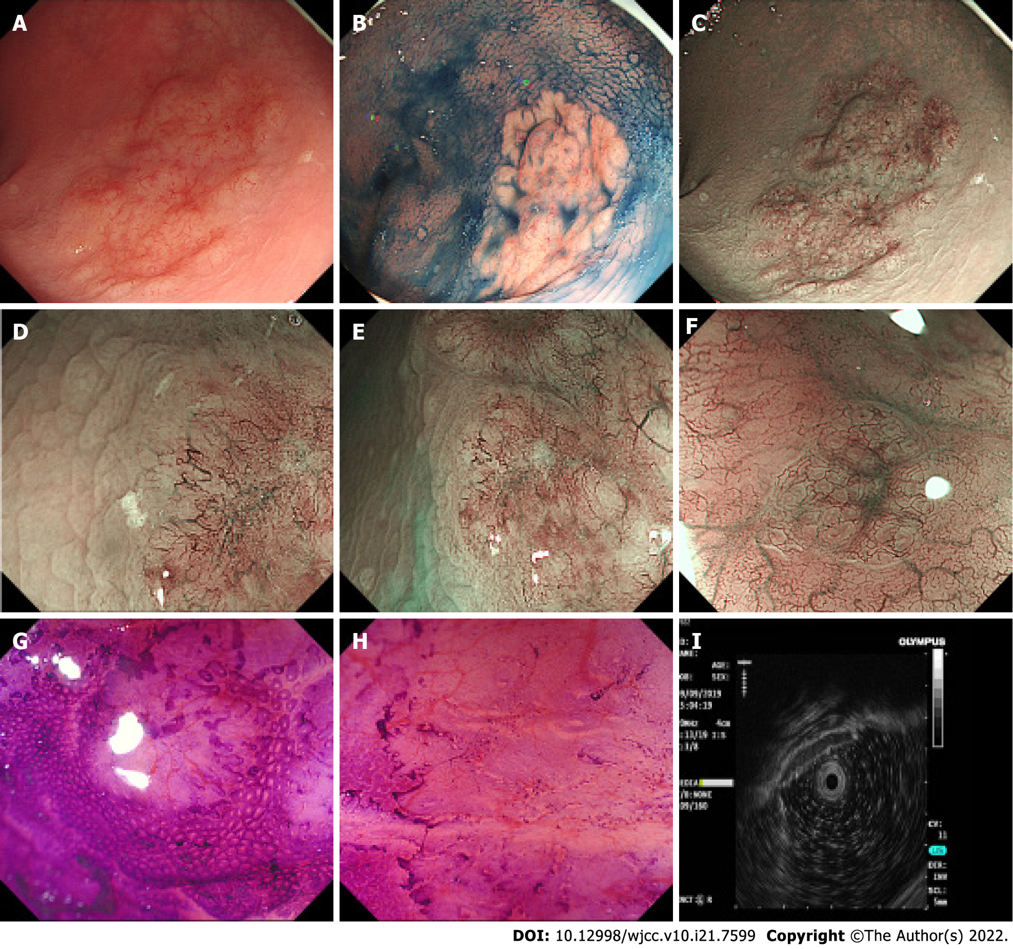
Figure 1 Endoscopic findings.
A: A 3.0 cm x 3.5 cm laterally spreading tumour-like elevated lesion was observed in the rectum; B: Indigo carmine staining showed a clear margin and uneven surface; C-F: Narrow band imaging (C) and magnifying endoscopy (D, E, F) showed a dark brown background and enlarged branch-like vessels on the surface of the lesion; G and H: Crystal violet staining magnifying endoscopy showed that the pit pattern structure disappeared on the surface of the lesion; I: Hypoechoic thickening of the mucosal layer was detected by endoscopic ultrasound.
- Citation: Tao Y, Nan Q, Lei Z, Miao YL, Niu JK. Rare primary rectal mucosa-associated lymphoid tissue lymphoma with curative resection by endoscopic submucosal dissection: A case report and review of literature. World J Clin Cases 2022; 10(21): 7599-7608
- URL: https://www.wjgnet.com/2307-8960/full/v10/i21/7599.htm
- DOI: https://dx.doi.org/10.12998/wjcc.v10.i21.7599


