Copyright
©The Author(s) 2022.
World J Clin Cases. Jul 26, 2022; 10(21): 7571-7576
Published online Jul 26, 2022. doi: 10.12998/wjcc.v10.i21.7571
Published online Jul 26, 2022. doi: 10.12998/wjcc.v10.i21.7571
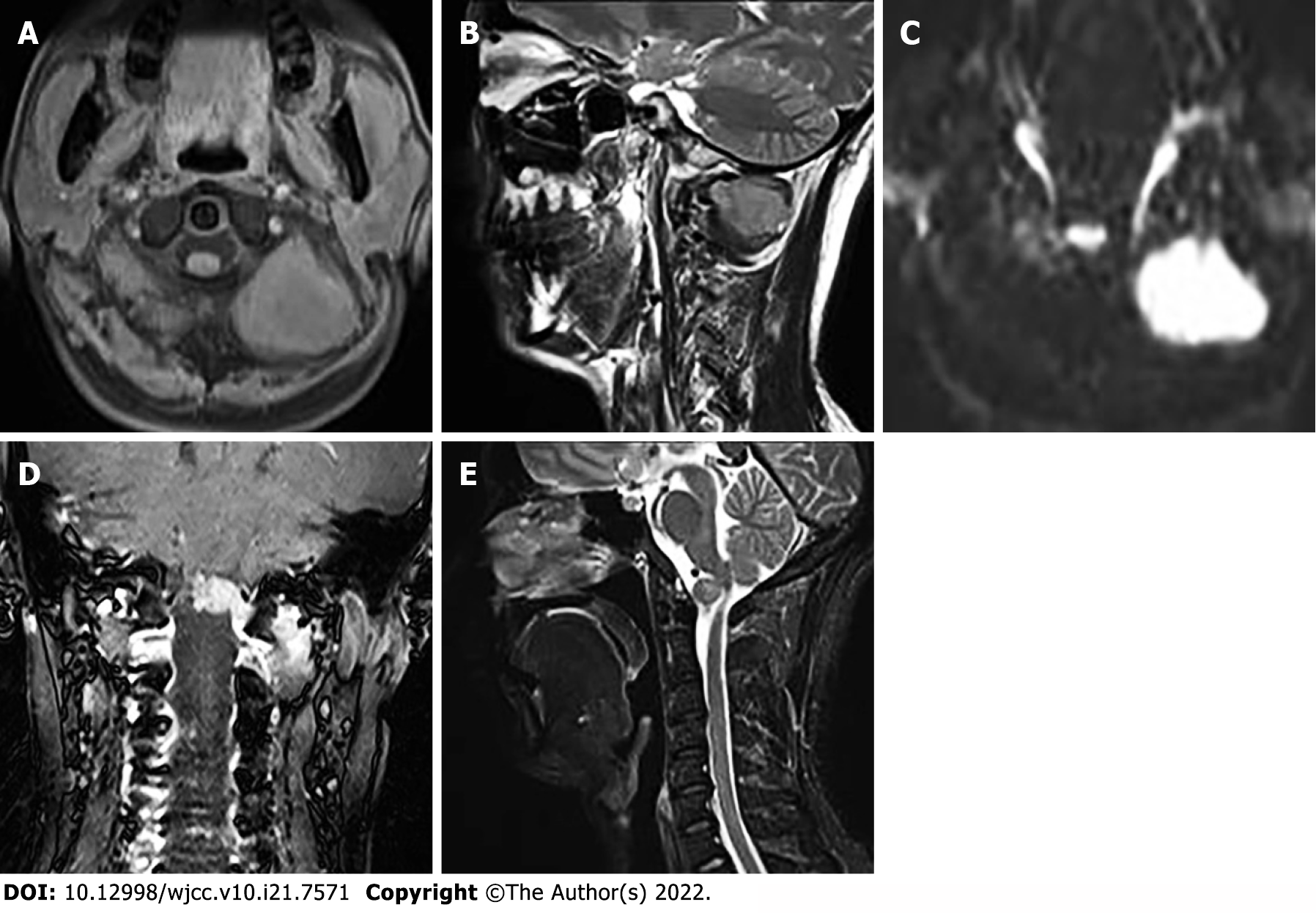
Figure 2 Magnetic resonance imaging of the neck.
A: Iso-signal on axial T1-weighted image; B: Slightly higher signal on sagittal T2-weighted image (T2WI); C: Significantly high signal on diffusion weighted imaging; D: Invasion of the anterior medulla oblongata with obvious uneven enhancement; E: The protrusion of the mass locally compressed the cervical spinal cord on T2WI.
- Citation: Liu CC, Huang WP, Gao JB. Primary clear cell sarcoma of soft tissue in the posterior cervical spine invading the medulla oblongata: A case report. World J Clin Cases 2022; 10(21): 7571-7576
- URL: https://www.wjgnet.com/2307-8960/full/v10/i21/7571.htm
- DOI: https://dx.doi.org/10.12998/wjcc.v10.i21.7571


