Copyright
©The Author(s) 2022.
World J Clin Cases. Jul 26, 2022; 10(21): 7571-7576
Published online Jul 26, 2022. doi: 10.12998/wjcc.v10.i21.7571
Published online Jul 26, 2022. doi: 10.12998/wjcc.v10.i21.7571
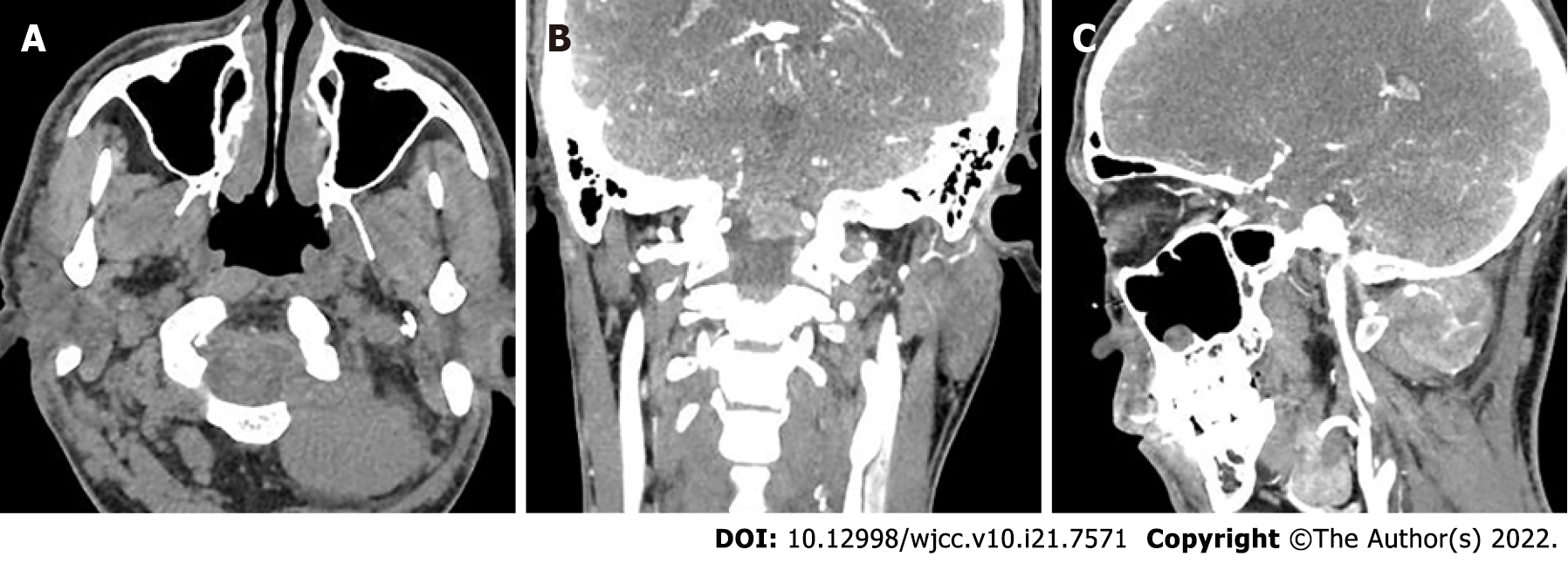
Figure 1 Computed tomography angiography images showing a posterior cervical soft tissue mass.
A: Plain scanning (axial view) showed a left paravertebral soft tissue mass at 1-2 levels in the neck, with clear boundaries; B: Enhanced scanning (coronal view) showed a mass protruding into the spinal canal of the cranio-cervical junction through the foramen magnum; C: Enhanced scanning (sagittal view) showed that the supplying artery was tortuous.
- Citation: Liu CC, Huang WP, Gao JB. Primary clear cell sarcoma of soft tissue in the posterior cervical spine invading the medulla oblongata: A case report. World J Clin Cases 2022; 10(21): 7571-7576
- URL: https://www.wjgnet.com/2307-8960/full/v10/i21/7571.htm
- DOI: https://dx.doi.org/10.12998/wjcc.v10.i21.7571


