Copyright
©The Author(s) 2022.
World J Clin Cases. Jul 26, 2022; 10(21): 7523-7530
Published online Jul 26, 2022. doi: 10.12998/wjcc.v10.i21.7523
Published online Jul 26, 2022. doi: 10.12998/wjcc.v10.i21.7523
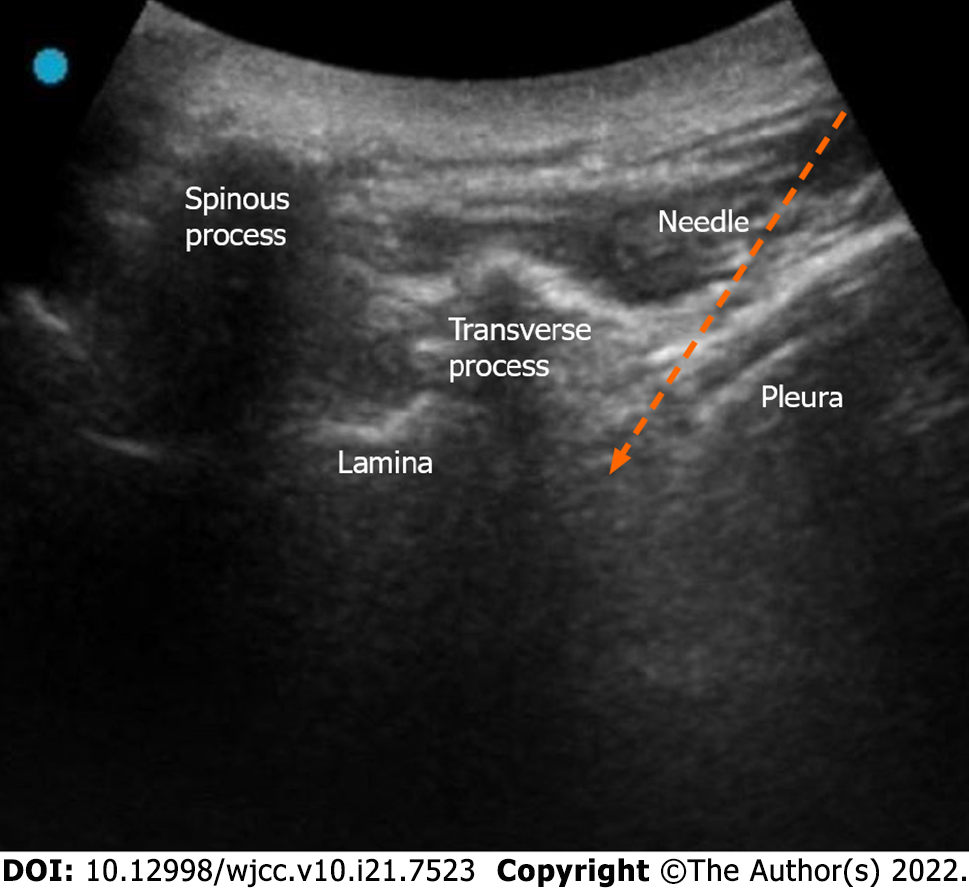
Figure 5 Ultrasound image of the selective nerve block.
Low-frequency probe short axis image showing the structure of spinous process, lamina, transverse process, and pleura from medial to lateral. The gap between the pleura and the lateral deep surface of the transverse process was the target region of the puncture (arrow).
- Citation: Zhang X, Li ZX, Yin LJ, Chen H. Selective nerve block for the treatment of neuralgia in Kummell’s disease: A case report. World J Clin Cases 2022; 10(21): 7523-7530
- URL: https://www.wjgnet.com/2307-8960/full/v10/i21/7523.htm
- DOI: https://dx.doi.org/10.12998/wjcc.v10.i21.7523


