Copyright
©The Author(s) 2022.
World J Clin Cases. Jul 26, 2022; 10(21): 7523-7530
Published online Jul 26, 2022. doi: 10.12998/wjcc.v10.i21.7523
Published online Jul 26, 2022. doi: 10.12998/wjcc.v10.i21.7523
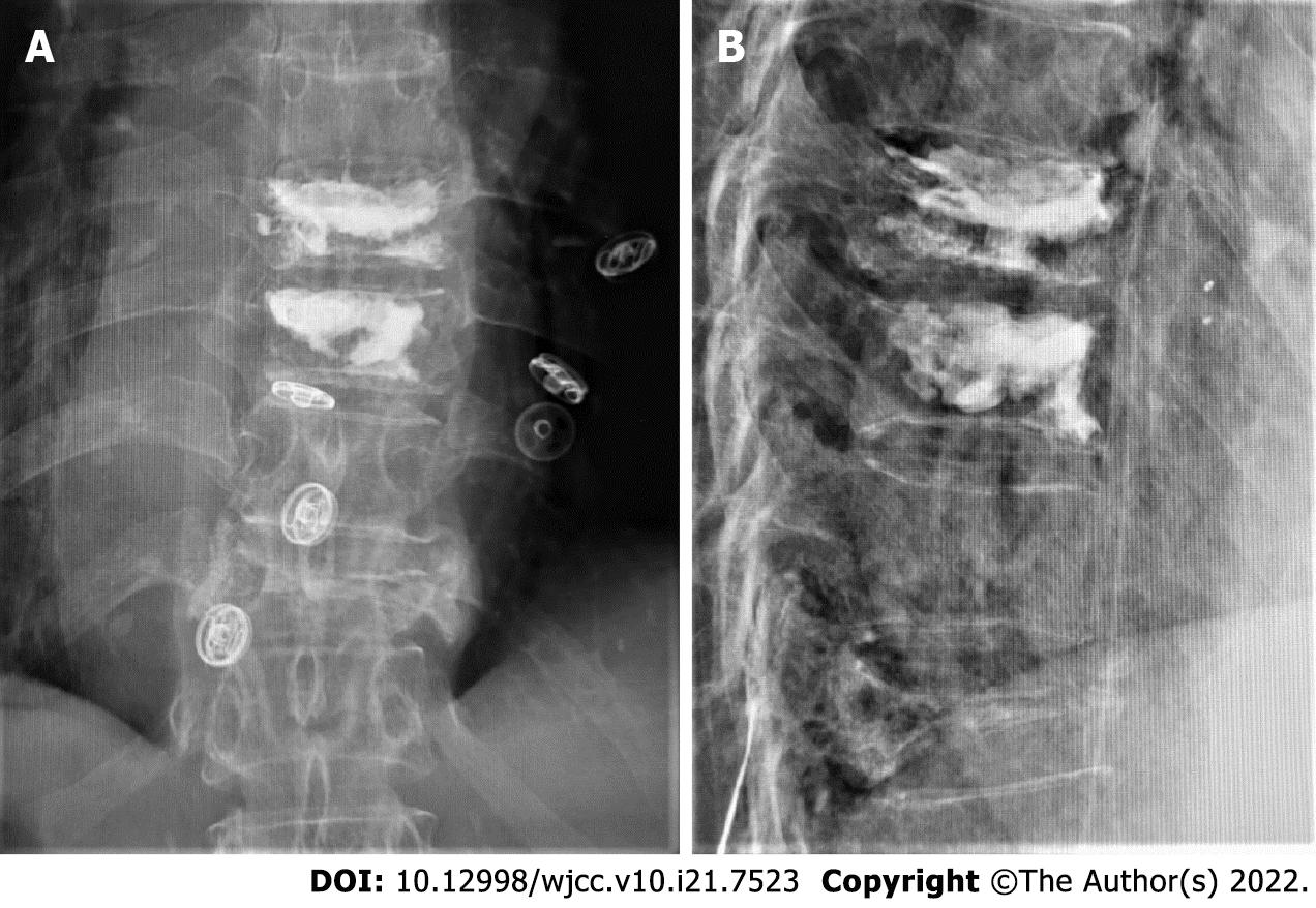
Figure 4 Postoperative radiograph of the patient.
A: Anterior-posterior film. B: Lateral film. The X-ray images show good distribution of the bone cement without marked leakage in the canalis spinalis.
- Citation: Zhang X, Li ZX, Yin LJ, Chen H. Selective nerve block for the treatment of neuralgia in Kummell’s disease: A case report. World J Clin Cases 2022; 10(21): 7523-7530
- URL: https://www.wjgnet.com/2307-8960/full/v10/i21/7523.htm
- DOI: https://dx.doi.org/10.12998/wjcc.v10.i21.7523


