Copyright
©The Author(s) 2022.
World J Clin Cases. Jul 26, 2022; 10(21): 7509-7516
Published online Jul 26, 2022. doi: 10.12998/wjcc.v10.i21.7509
Published online Jul 26, 2022. doi: 10.12998/wjcc.v10.i21.7509
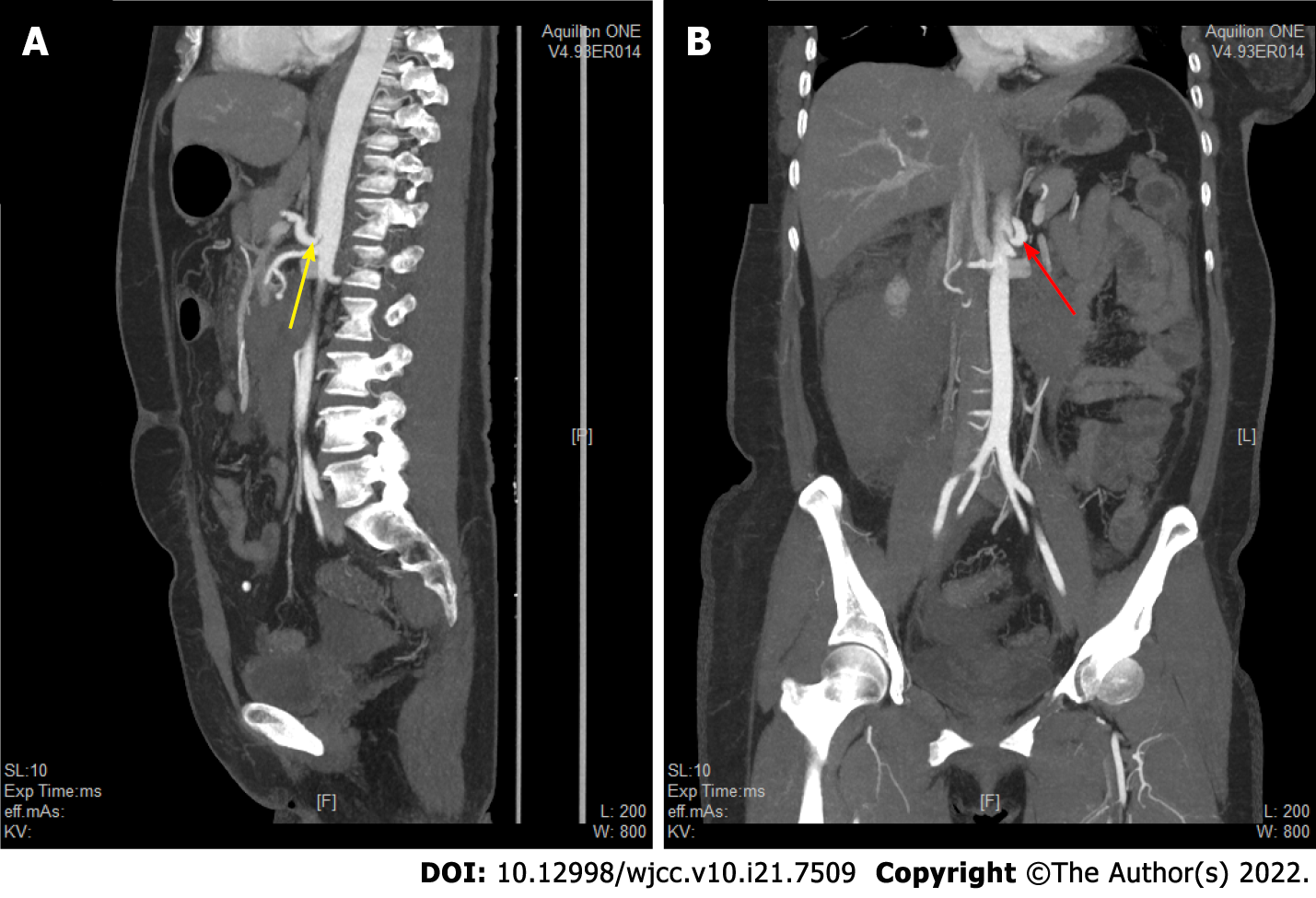
Figure 4 Abdominal enhanced computed tomography examination showed that the abdominal cavity was narrow.
A: Sagittal image (as shown by the yellow arrow) showing that the abdominal cavity aortic opening is at the T12 level. The starting point of the celiac trunk is narrow and abnormal, and the upper edge of the proximal wall of the celiac trunk is a sharp “V-shaped” depression; B: Coronal image (as shown by the red arrow) showing abnormal stenosis at the origin of the celiac trunk.
- Citation: Lu XC, Pei JG, Xie GH, Li YY, Han HM. Median arcuate ligament syndrome with retroperitoneal haemorrhage: A case report. World J Clin Cases 2022; 10(21): 7509-7516
- URL: https://www.wjgnet.com/2307-8960/full/v10/i21/7509.htm
- DOI: https://dx.doi.org/10.12998/wjcc.v10.i21.7509


