Copyright
©The Author(s) 2022.
World J Clin Cases. Jul 26, 2022; 10(21): 7502-7508
Published online Jul 26, 2022. doi: 10.12998/wjcc.v10.i21.7502
Published online Jul 26, 2022. doi: 10.12998/wjcc.v10.i21.7502
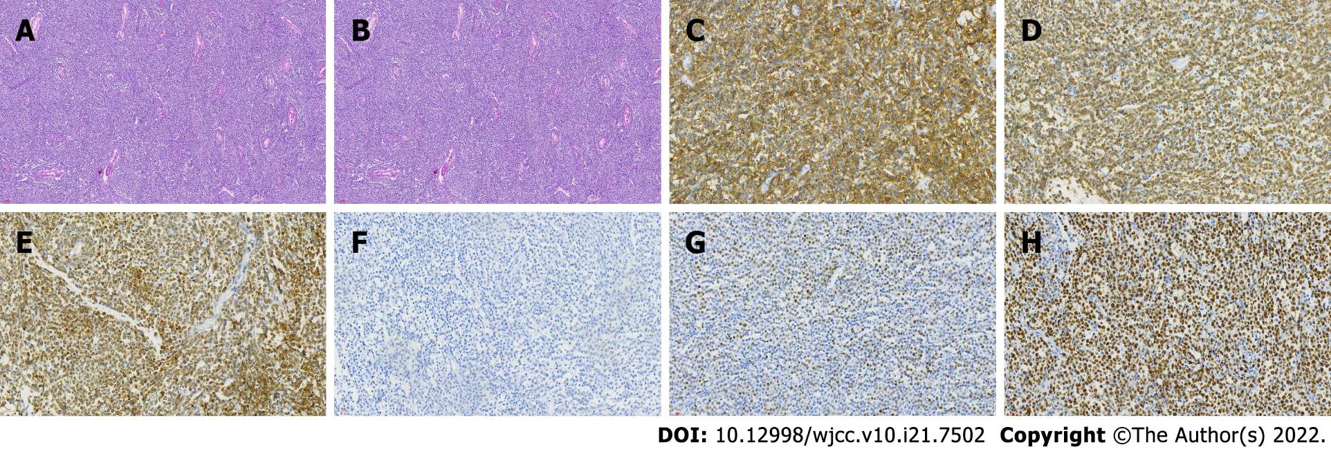
Figure 1 Histopathological microphotograph of primary testicular diffuse large B-cell lymphoma.
A: The basic structure of the testicle is destroyed, and there are a lot of lymphocytes infiltrating (magnification × 100); B: Hematoxylin and eosin stained sections showed diffuse proliferation of medium-sized round cells (magnification × 400); C-H: On immunohistochemistry, the neoplastic cells showed positive expression of CD20 (C), CD79a (D), BCL-2 (E), BCL-6 (F), C-myc (G); Ki67 staining showed almost 80% proliferation index (H).
- Citation: Zhang CJ, Zhang JY, Li LJ, Xu NW. Refractory lymphoma treated with chimeric antigen receptor T cells combined with programmed cell death-1 inhibitor: A case report. World J Clin Cases 2022; 10(21): 7502-7508
- URL: https://www.wjgnet.com/2307-8960/full/v10/i21/7502.htm
- DOI: https://dx.doi.org/10.12998/wjcc.v10.i21.7502


